. 2010 Sep 8;30(36):12210-8.
doi: 10.1523/JNEUROSCI.1520-10.2010.

Tumor necrosis factor-alpha (TNF-alpha) regulates shedding of TNF-alpha receptor 1 by the metalloprotease-disintegrin ADAM8: evidence for a protease-regulated feedback loop in neuroprotection

Affiliations

Tumor necrosis factor-alpha (TNF-alpha) regulates shedding of TNF-alpha receptor 1 by the metalloprotease-disintegrin ADAM8: evidence for a protease-regulated feedback loop in neuroprotection

Jörg W Bartsch et al. J Neurosci. .

Abstract

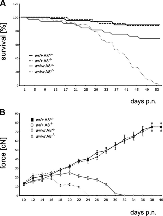
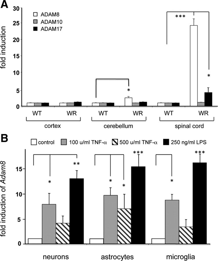
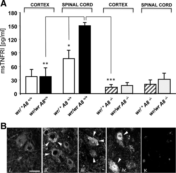
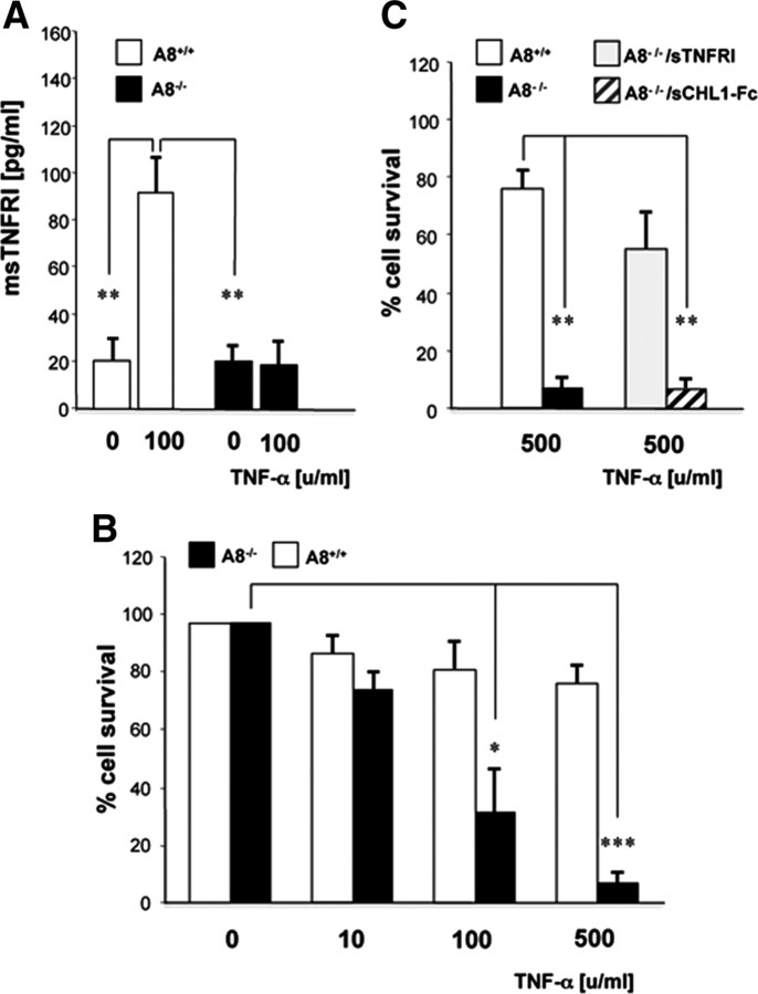
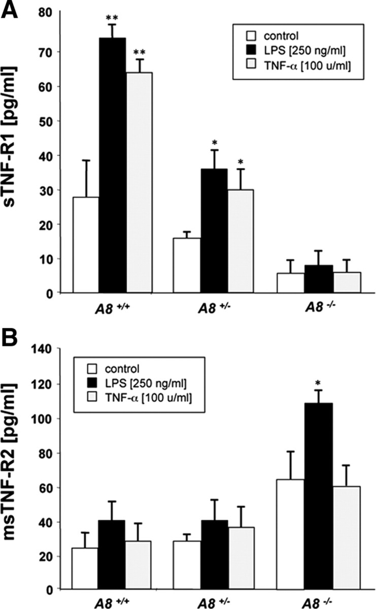
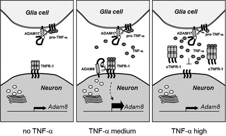
Tumor necrosis factor alpha (TNF-alpha) is a potent cytokine in neurodegenerative disorders, but its precise role in particular brain disorders is ambiguous. In motor neuron (MN) disease of the mouse, exemplified by the model wobbler (WR), TNF-alpha causes upregulation of the metalloprotease-disintegrin ADAM8 (A8) in affected brain regions, spinal cord, and brainstem. The functional role of A8 during MN degeneration in the wobbler CNS was investigated by crossing WR with A8-deficient mice: a severely aggravated neuropathology was observed for A8-deficient WR compared with WR A8(+/-) mice, judged by drastically reduced survival [7 vs 81% survival at postnatal day 50 (P50)], accelerated force loss in the forelimbs, and terminal akinesis. In vitro protease assays using soluble A8 indicated specific cleavage of a TNF-alpha receptor 1 (p55 TNF-R1) but not a TNF-R2 peptide. Cleavage of TNF-R1 was confirmed in situ, because levels of soluble TNF-R1 were increased in spinal cords of standard WR compared with wild-type mice but not in A8-deficient WR mice. In isolated primary neurons and microglia, TNF-alpha-induced TNF-R1 shedding was dependent on the A8 gene dosage. Furthermore, exogenous TNF-alpha showed higher toxicity for cultured neurons from A8-deficient than for those from wild-type mice, demonstrating that TNF-R1 shedding by A8 is neuroprotective. Our results indicate an essential role for ADAM8 in modulating TNF-alpha signaling in CNS diseases: a feedback loop integrating TNF-alpha, ADAM8, and TNF-R1 shedding as a plausible mechanism for TNF-alpha mediated neuroprotection in situ and a rationale for therapeutic intervention.


Figures

Figure 1.
Figure 1.
Absence of ADAM8 aggravates the wobbler (WR) disease. A, Kaplan–Meier survival curves of wr/+ Adam8 +/+ (black line; n = 45), wr/+ Adam8 −/− (dashed black line; n = 34), wr/wr Adam8 +/− (gray line; n = 32), and wr/wr Adam8 −/− (dashed gray line; n = 38) mice for 54 d after birth. Significance was calculated by pairwise log rank tests for survival with p < 0.07. B, Grip strengths of forelimbs of wr/+ Adam8 +/+ (squares), wr/+ Adam8 −/− (circles), wr/wr Adam8 +/− (rhombs), and wr/wr Adam8 −/− (triangles) mice. For all genotypes, n = 15. Mean ± SD values. Note the total loss of force within 24 d after birth in wr/wr Adam8 −/− individuals, leading to complete akinesis and premature death. p.n., Postnatal.
Figure 2.
Figure 2.
WR histopathology of spinal cord is aggravated by ADAM8 deficiency. A–C, Morphology of spinal cord neurons in the anterior horn of mice with wr/+ Adam8 −/− (A), wr/wr Adam8 +/− (B), and wr/wr Adam8 −/− (C) genotypes, respectively. Spinal cord sections were stained with cresyl violet and immunohistochemistry/DAB staining (brown) for microglia (CD45). The ventrolateral part of the spinal cord is shown (white matter to the left in A and to the right in B and C). Motor neurons in A show normal morphology (black arrows). Motor neurons in wr/wr Adam8 +/− mice (B) display early stages of degeneration, characterized by chromatolysis and fading, characteristic features of neurodegeneration in WR mice (asterisks). In addition, compared with A, increased staining of small CD45-positive microglia is detectable surrounding degenerating motor neurons (white arrows in B). In C, increased numbers of degenerated neurons in the spinal cord of wr/wr A8 −/− compared with wr/wr Adam8 +/− mice (black arrows, unaffected neuron) and degenerating neurons of different stages (* and ** reflect different stages of degeneration, respectively). D, E, More detailed views of degenerating motor neurons in wr/wr Adam8 +/− and wr/wr A8 −/− WR mice with additional vacuolized motor neurons (E). There were no signs of morphological differences between degeneration observed in wr/wr Adam8 +/− and wr/wr A8 −/− spinal cords. In F–H, immunohistochemistry for GFAP (reactive gliosis) and CD45 immunostaining for microglia activation (I–M) for genotypes wr/+ Adam8 −/− (I), wr/wr Adam8 +/− (J, L), and wr/wr Adam8 −/− (K, M) genotypes. L, M, Enlarged view of microglia indicated by arrowheads in J and K, respectively. Note the chain-like appearance of microglia in wr/wr A8 −/− mice, resulting from lesser ramification. Scale bars: (in A) A–C, 50 μm; (in D) D, E, 10 μm; (in F) F–K, 50 μm; (in L) L, M, 10 μm.
Figure 3.
Figure 3.
Quantitative evaluation of histopathological data. A, Motor neuron numbers in ventrolateral pool of anterior horns, judged by Nissl staining; B, GFAP immunoreactivity, given as pixel values per spinal cord section; C, quantification of CD45 immunoreactivity. In each bar graph, quantification of image data were performed for wr/+ Adam8 −/− (black bars), wr/wr Adam8 +/− (gray bars), and wr/wr Adam8 −/− (white bars). Pixel values from individual images were acquired using NIH ImageJ analysis software. *p < 0.05, ***p < 0.001.
Figure 4.
Figure 4.
ADAM mRNA levels in WR mice and in primary CNS cells. Levels of ADAM8, ADAM10, and ADAM17 mRNA in cortex, cerebellum, and spinal cords of WT and WR mice are elevated by neurodegeneration (wobbler CNS), LPS, and TNF-α in neurons, astrocytes, and microglia. A, Quantitative RT-PCR analysis of Adam genes encoding ADAM8, ADAM10, and ADAM17 in the CNS of WT and WR cortex, cerebellum, and spinal cord. B, Quantitative RT-PCR analysis for Adam8 in primary neurons (cerebellar granular cells), spinal cord astrocytes, and cortical microglia cells from mouse CNS in the presence of recombinant mouse TNF-α (100 ng/ml) or LPS (250 ng/ml). Light cycler data were normalized to reference mRNAs (Ubiquitin C and ribosomal protein L7) and given relative to WT. Mean ± SD values were derived by three independent runs performed in triplets for each sample (n = 9). *p < 0.05, **p < 0.01, ***p < 0.001.
Figure 5.
Figure 5.
A, Concentrations of soluble TNF-R1 in the spinal cord of wobbler mice determined by ELISA. Lysates from cortex and spinal cord from wild-type Adam8 +/+ wr/+ (white bars), Adam8 +/+ wr/wr (black bars), Adam8 −/− wr/+ (hatched bars), and Adam8 −/− wr/wr (gray bars) were analyzed with an sTNF-R1 ELISA kit. Mean ± SD values from three independent experiments performed in triplets (n = 9). Data were analyzed by t test. *p < 0.05, **p < 0.01, ***p < 0.001. B, Immunohistochemistry of TNF-R1 in spinal cord sections corresponding to the samples analyzed in A: i, wr/+ Adam8 +/+ with large intact motor neurons, weakly stained for TNF-R1; ii, wr/+ Adam8 −/− with vesicular and membrane-associated TNF-R1 signals; iii, wr/wr Adam8 +/− increased vesicular TNF-R1 staining compared with ii; iv, wr/wr Adam8 −/− with strong TNF-R1 staining throughout the cell body; v, negative control (omission of primary antibody). Scale bar, 50 μm. Immunostaining for TNF-R1 is indicated by white arrowheads.
Figure 6.
Figure 6.
Effect of TNF-α on cultured cerebellar neurons from wild-type and Adam8-deficient mice. A, TNF-α (10 ng/ml = 100 U/ml) induces the release of soluble TNF-R1 from A8+/+ neurons (white bars), which is dependent on ADAM8, because there is no induction in ADAM8-deficient neurons (black bars). In B, ADAM8-deficient neurons show a higher sensitivity to TNF-α-induced cell death. Less than 10% of the cells survive a TNF-α dosage of 500 ng/ml, whereas >80% of wild-type cells survived. In C, TNF-α (100 U/ml)-induced neuronal cell death was prevented by incubation with an excess of soluble TNF-R1 (10 ng/ml) but not with soluble recombinant CHL1 (sCHL1-Fc; 50 ng/ml), a neuronal substrate of ADAM8 that was shown to protect neurons from constitutive cell death. *p < 0.05, **p < 0.01, ***p < 0.001.
Figure 7.
Figure 7.
A, B, Release of soluble TNF-R1 (A) and TNF-R2 (B) from primary microglia cells isolated from wild-type, heterozygous, and homozygous Adam8 knock-out mice and induced with indicated concentrations of LPS or TNF-α. ELISA data (in picograms per milliliter medium) were acquired from three independent experiments performed in triplets (n = 9) and were given as mean ± SD. *p < 0.05, **p < 0.01.
Figure 8.
Figure 8.
Proposed neuroprotective feedback loop involving TNF-α, TNF-R1, and ADAM8. At low TNF-α concentrations, the transcription rate of the Adam8 gene is low and very low levels of soluble TNF-R1 are found in the extracellular compartment. At high TNF-α concentrations (e.g., 100 U/ml), TNF-R1 activates intracellular pathway(s) strongly stimulating transcription of the Adam8 gene. Its product, the cell-surface-exposed protease ADAM8 cleaves the transmembrane protein TNF-R1 and releases sTNF-R1 from the cell into the intercellular space, in which high concentrations of sTNF-R1 are able to scavenge free TNF-α. This in turn reduces the cellular response to TNF-α, i.e., preventing activation of TNF-dependent genes involved in neuronal cell death.

References

    1. Bigini P, Repici M, Cantarella G, Fumagalli E, Barbera S, Cagnotto A, De Luigi A, Tonelli R, Bernardini R, Borsello T, Mennini T. Recombinant human TNF-binding protein-1 (rhTBP-1) treatment delays both symptoms progression and motor neuron loss in the wobbler mouse. Neurobiol Dis. 2008;29:465–476. - PubMed
    1. Black RA. Tumor necrosis factor-alpha converting enzyme. Int J Biochem Cell Biol. 2002;34:1–5. - PubMed
    1. Black RA, Rauch CT, Kozlosky CJ, Peschon JJ, Slack JL, Wolfson MF, Castner BJ, Stocking KL, Reddy P, Srinivasan S, Nelson N, Boiani N, Schooley KA, Gerhart M, Davis R, Fitzner JN, Johnson RS, Paxton RJ, March CJ, Cerretti DP. A metalloproteinase disintegrin that releases tumour-necrosis factor-alpha from cells. Nature. 1997;385:729–733. - PubMed
    1. Chen G, Goeddel DV. TNF-R1 signaling: a beautiful pathway. Science. 2002;296:1634–1635. - PubMed
    1. Cui X, Hawari F, Alsaaty S, Lawrence M, Combs CA, Geng W, Rouhani FN, Miskinis D, Levine SJ. Identification of ARTS-1 as a novel TNFR1-binding protein that promotes TNFR1 ectodomain shedding. J Clin Invest. 2002;110:515–526. - PMC - PubMed

Publication types

MeSH terms