. 2010 Nov 5;285(45):35206-15.
doi: 10.1074/jbc.M110.171769. Epub 2010 Sep 8.

Plasma kallikrein promotes epidermal growth factor receptor transactivation and signaling in vascular smooth muscle through direct activation of protease-activated receptors

Affiliations

Plasma kallikrein promotes epidermal growth factor receptor transactivation and signaling in vascular smooth muscle through direct activation of protease-activated receptors

Rany T Abdallah et al. J Biol Chem. .

Erratum in

  • J Biol Chem. 2011 Jul 1;286(26):23620. El-Shewy, Hesham M [added]

Abstract

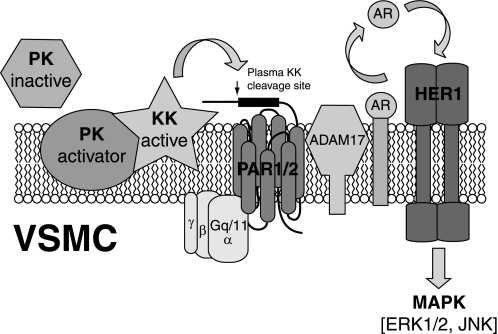
The kallikrein-kinin system, along with the interlocking renin-angiotensin system, is a key regulator of vascular contractility and injury response. The principal effectors of the kallikrein-kinin system are plasma and tissue kallikreins, proteases that cleave high molecular weight kininogen to produce bradykinin. Most of the cellular actions of kallikrein (KK) are thought to be mediated by bradykinin, which acts via G protein-coupled B1 and B2 bradykinin receptors on VSMCs and endothelial cells. Here, we find that primary aortic vascular smooth muscle but not endothelial cells possess the ability to activate plasma prekallikrein. Surprisingly, exposing VSMCs to prekallikrein leads to activation of the ERK1/2 mitogen-activated protein kinase cascade via a mechanism that requires kallikrein activity but does not involve bradykinin receptors. In transfected HEK293 cells, we find that plasma kallikrein directly activates G protein-coupled protease-activated receptors (PARs) 1 and 2, which possess consensus kallikrein cleavage sites, but not PAR4. In vascular smooth muscles, KK stimulates ADAM (a disintegrin and metalloprotease) 17 activity via a PAR1/2 receptor-dependent mechanism, leading sequentially to release of the endogenous ADAM17 substrates, amphiregulin and tumor necrosis factor-α, metalloprotease-dependent transactivation of epidermal growth factor receptors, and metalloprotease and epidermal growth factor receptor-dependent ERK1/2 activation. These results suggest a novel mechanism of bradykinin-independent kallikrein action that may contribute to the regulation of vascular responses in pathophysiologic states, such as diabetes mellitus.


Figures

FIGURE 1.
FIGURE 1.
Activation of plasma prekallikrein in primary vascular smooth muscle but not endothelial cells. Serum-deprived R-VSMCs, H-VSMCs, human umbilical vein endothelial cells (HUVEC), or human aortic endothelial cells (HAEC) in 96-well plates were incubated with 100 nm human PK in the presence of the chromogenic substrate for plasma KK, S2302 (0.6 mm), and KK activity was quantified as change in absorption at 405 nm measured at 2-min intervals over 2 h. Data shown are from one of three experiments, which produced comparable results.
FIGURE 2.
FIGURE 2.
Activation of ERK1/2 in primary rat aortic vascular smooth muscle cells by prekallikrein is independent of bradykinin receptors. A, serum-deprived R-VSMCs in six-well plates were treated with human plasma PK at increasing concentrations for 15 min (left panel) or with 200 nm plasma PK for varying times (right panel) and ERK1/2 phosphorylation was determined by immunoblotting whole cell lysates as described. Total cell ERK1/2 was blotted as a control for equal protein loading. B, serum-deprived R-VSMCs in six-well plates were preincubated for 15 min in the presence or absence of the BK receptor antagonist, HOE140 (10 μm; Sigma), prior to stimulation with 10 nm BK (left panel) or 200 nm human plasma PK (right panel) for 15 min. ERK1/2 phosphorylation was determined from whole cell lysates as described. Total cell ERK1/2 was blotted as a control for equal protein loading. Each bar graph depicts the mean ± S.E. for three separate experiments. *, greater than untreated control, p < 0.05; †, greater than control, p < 0.01.
FIGURE 3.
FIGURE 3.
Plasma kallikrein directly activates PAR1 and PAR2 receptors. A, schematic depicting the N-terminal proteolytic cleavage sites and internal ligand sequences of PAR1–4. B, serum-deprived HEK293 cells expressing PAR1-GFP (P1) were treated for 15 min with vehicle (Veh), 1 nm thrombin (Thr) as a positive control, or 20 nm plasma KK, after which cells were fixed, permeabilized, co-stained with early endosome (EAA1) and nuclear markers, and examined under confocal fluorescence microscopy. The distribution of GFP-PAR1 (green), early endosomes (red), and cell nuclei (blue) are shown in individual channels along with a composite image. C, serum-deprived HEK293 cells expressing PAR1-GFP (P1), PAR2-GFP (P2), or PAR4-GFP (P4) were treated with 1 nm thrombin (Thr; PAR1/PAR4) or 100 μm trypsin (Trp; PAR2) for 15 min as positive controls or were treated with 20 nm plasma KK for 5 or 30 min, after which cells were fixed, and the distribution of GFP-PAR1/2/4 was examined under confocal fluorescence microscopy. Representative fields are shown from one of three experiments that produced comparable results. D, internalization of GFP-PAR1/2/4 in response to thrombin, trypsin, or KK was quantified by measuring the ratio of cytosolic GFP fluorescence to total cellular GFP fluorescence in confocal images of cells exposed to each condition, as described. The bar graph depicts the mean ± S.E. for three separate experiments. *, greater than untreated control, p < 0.05. NS, nonstimulated.
FIGURE 4.
FIGURE 4.
Plasma kallikrein causes PAR-dependent ADAM17 activation in vascular smooth muscle cells. A, serum-deprived R-VSMCs in 96-well plates were treated with varying concentrations of plasma KK for 4 h in the presence of the ADAM10/17-specific fluorogenic peptide substrate, MCA-KPLGL-Dpa-AR-NH2 (20 μm). ADAM activity was quantified as the increased fluorescence of the cleaved substrate and expressed as percent of the activity observed when the incubation was performed in the absence of KK. Data shown represent mean ± S.D. of triplicate determinations in one of three experiments that produced comparable results. *, greater than untreated control, p < 0.05. B, serum-deprived R-VSMCs in 10-cm plates were treated with 20 nm plasma KK for 5–15 min, after which cells were lysed, and ADAM17 activity was measured using the Innozyme TACE Activity KitTM as described. Data shown represent mean ± S.D. of triplicate determinations in one of three experiments that produced comparable results. *, greater than untreated control, p < 0.05. C, 20 nm human plasma KK was mixed with the chromogenic substrate for plasma KK, S2302 (0.6 mm), in the presence or absence of the PAR1 antagonist FLLRN (51) (P1Inh; 200 μm) or PAR2 antagonist FSLLRY (52) (P2Inh; 200 μm) (Peptides International; Louisville, KY), and KK activity was measured as the change in absorption measured at 405 nm. D, serum-deprived R-VSMCs in 96-well plates were preincubated for 15 min in the presence or absence of PAR1 or PAR2 antagonist peptides (200 μm) prior to treatment with 20 nm plasma KK for 4 h in the presence of MCA-KPLGL-Dpa-AR-NH2. ADAM activity was quantified as the increased fluorescence of the cleaved substrate and expressed as percent of the activity observed when the incubation was performed in the absence of KK. Data shown represent mean ± S.D. of triplicate determinations in one of three experiments that produced comparable results. *, greater than untreated control, p < 0.05. †, less than KK treated, p < 0.05.
FIGURE 5.
FIGURE 5.
Plasma kallikrein stimulates ADAM-dependent amphiregulin and TNF-α release from vascular smooth muscle cells. A, serum-deprived H-VSMC in 10-cm dishes were incubated in the presence or absence of 50 nm human plasma KK for 1–3 h, after which the medium was collected, concentrated, and immunoblotted for AR or TNFα, as described. B, serum-deprived H-VSMC were incubated with or without 50 nm human plasma KK in the presence or absence of GM6001 (10 μm; Calbiochem), and AR shedding was determined. Data are expressed as the percent increase in AR or TNF-α abundance compared with incubations performed in the absence of KK. Each bar graph depicts the mean ± S.E. for three separate experiments. *, greater than untreated control, p < 0.05; †, less than KK treated, p < 0.05. NS, nonstimulated.
FIGURE 6.
FIGURE 6.
Plasma kallikrein stimulates ADAM-dependent EGF receptor transactivation in vascular smooth muscle cells. A, 20 nm human plasma KK was combined with the chromogenic substrate for plasma KK, S2302 (0.6 μm), in the presence or absence of GM6001 (10 μm), and KK activity was measured as the change in absorption measured at 405 nm. B, serum-deprived R-VSMCs in six-well plates were preincubated in the presence or absence of GM6001 for 15 min prior to treatment with 20 nm plasma KK for 1–5 min. EGF receptor Tyr1068 phosphorylation was determined by immunoblotting whole cell lysates as described. Data shown represents the mean ± S.E. for five separate experiments. *, greater than untreated control, p < 0.05; †, less than KK treated, p < 0.05. NS, nonstimulated (vehicle).
FIGURE 7.
FIGURE 7.
Activation of ERK1/2 by plasma kallikrein in vascular smooth muscle cells is partially ADAM- and EGF receptor-dependent. A, serum-deprived R-VSMCs in 6-well plates were preincubated in the presence or absence of GM6001 (10 μm) for 15 min prior to treatment with 200 nm PK or 20 nm plasma KK for 15 min. ERK1/2 phosphorylation was determined by immunoblotting whole cell lysates as described. Total cell ERK1/2 was blotted as a control for equal protein loading. B, serum-deprived R-VSMCs were preincubated in the presence or absence of GM6001 for 15 min prior to treatment with 200 nm PK or 20 nm plasma KK for 15 min. JNK phosphorylation was determined by immunoblotting whole cell lysates, and total cell JNK was blotted as a control for equal protein loading. C, serum-deprived R-VSMCs were preincubated in the presence or absence of AG1478 (100 nm) for 15 min prior to treatment with 10 nm EGF or 20 nm plasma KK for 15 min. ERK1/2 phosphorylation was determined by immunoblotting whole cell lysates, and total cell ERK1/2 was blotted as a control for equal protein loading. Each bar graph represents the mean ± S.E. for three separate experiments. *, greater than untreated control, p < 0.05; †, less than stimulated in the absence of inhibitor, p < 0.05. NS, nonstimulated (vehicle).
FIGURE 8.
FIGURE 8.
Proposed mechanism of bradykinin-independent kallikrein effects in vascular smooth muscle cells. Circulating plasma PK is activated to KK upon binding to the surface of exposed VSMC. The N termini of PAR1 and PAR2 undergo KK-dependent cleavage, exposing their internal tethered ligands and promoting PAR-dependent activation of ADAM17. ADAM17 activation releases AR, causing EGF receptor transactivation and EGF receptor-dependent activation of downstream signaling pathways including the ERK and JNK cascades.

References

    1. Sainz I. M., Pixley R. A., Colman R. W. (2007) Thromb. Haemost. 98, 77–83 - PubMed
    1. Hermann A., Arnhold M., Kresse H., Neth P., Fink E. (1999) Immunopharmacology 45, 135–139 - PubMed
    1. Schmaier A. H. (2008) Int. Immunopharmacol. 8, 161–165 - PMC - PubMed
    1. Motta G., Rojkjaer R., Hasan A. A., Cines D. B., Schmaier A. H. (1998) Blood 91, 516–528 - PubMed
    1. Motta G., Shariat-Madar Z., Mahdi F., Sampaio C. A., Schmaier A. H. (2001) Thromb. Haemost. 86, 840–847 - PubMed

Publication types

MeSH terms

Substances