. 2010 Nov 12;285(46):35606-14.
doi: 10.1074/jbc.M110.142406. Epub 2010 Sep 7.

Sustained glutamate receptor activation down-regulates GABAB receptors by shifting the balance from recycling to lysosomal degradation

Affiliations

Sustained glutamate receptor activation down-regulates GABAB receptors by shifting the balance from recycling to lysosomal degradation

Patrick J Maier et al. J Biol Chem. .

Abstract

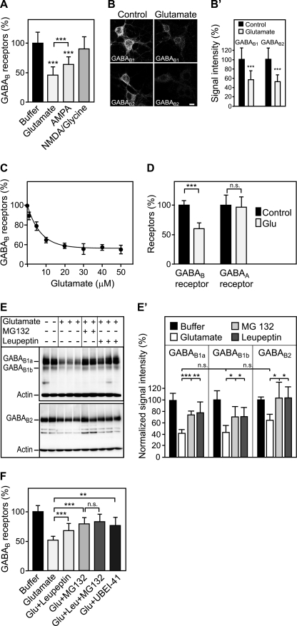
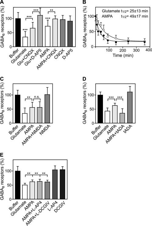
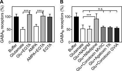
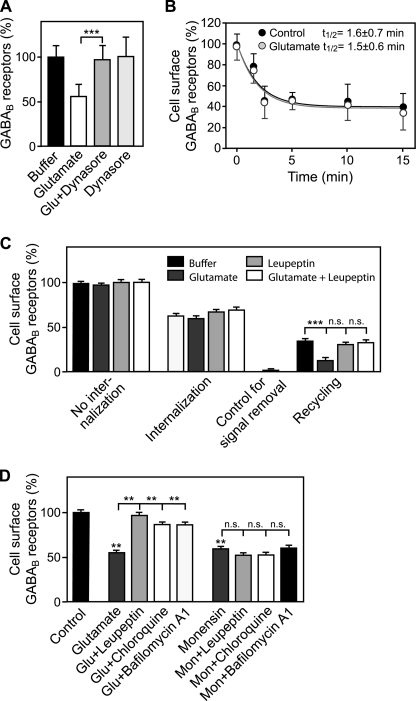
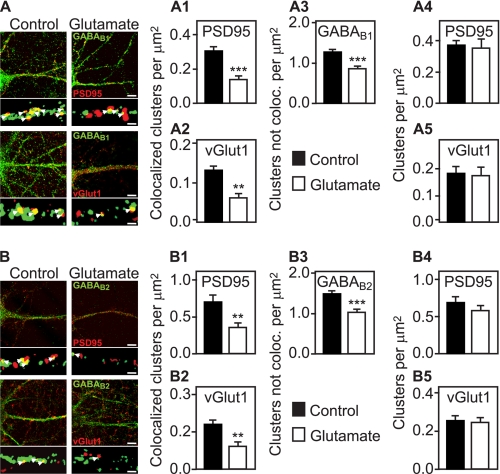
Metabotropic GABA(B) receptors are abundantly expressed at glutamatergic synapses where they control excitability of the synapse. Here, we tested the hypothesis that glutamatergic neurotransmission may regulate GABA(B) receptors. We found that application of glutamate to cultured cortical neurons led to rapid down-regulation of GABA(B) receptors via lysosomal degradation. This effect was mimicked by selective activation of AMPA receptors and further accelerated by coactivation of group I metabotropic glutamate receptors. Inhibition of NMDA receptors, blockade of L-type Ca(2+) channels, and removal of extracellular Ca(2+) prevented glutamate-induced down-regulation of GABA(B) receptors, indicating that Ca(2+) influx plays a critical role. We further established that glutamate-induced down-regulation depends on the internalization of GABA(B) receptors. Glutamate did not affect the rate of GABA(B) receptor endocytosis but led to reduced recycling of the receptors back to the plasma membrane. Blockade of lysosomal activity rescued receptor recycling, indicating that glutamate redirects GABA(B) receptors from the recycling to the degradation pathway. In conclusion, the data indicate that sustained activation of AMPA receptors down-regulates GABA(B) receptors by sorting endocytosed GABA(B) receptors preferentially to lysosomes for degradation on the expense of recycling. This mechanism may relieve glutamatergic synapses from GABA(B) receptor-mediated inhibition resulting in increased synaptic excitability.


Figures

FIGURE 1.
FIGURE 1.
Sustained activation of glutamate receptors induces the down-regulation of GABAB receptors via lysosomal degradation. A, down-regulation of GABAB receptors is triggered by glutamate and AMPA. Primary cortical neurons were treated either with 50 μm glutamate, 100 μm AMPA, or 100 μm NMDA + 10 μm glycine for 90 min and tested for GABAB receptor levels using the in-cell Western assay and antibodies directed against the C terminus of GABAB1. Means ± S.D.; n = 20–40 cultures from three to five preparations; ***, p < 0.001, one-way ANOVA; Bonferoni post test. B, glutamate down-regulates GABAB1 and GABAB2 as verified by immunocytochemistry. For immunofluorescence analysis, cells were treated with 50 μm glutamate for 30 min. This incubation time resulted in sufficient remaining fluorescence signals in the glutamate-treated cultures enabling a reliable quantification. After fixation and permeabilization, neurons were stained with antibodies directed against GABAB1 and GABAB2, respectively. Representative confocal images are shown. B′, quantification of fluorescence signals. Scale bar represents 10 μm. Means ± S.D.; n = 33–48 neurons from three preparations; ***, p < 0.0001, t test. C, dose dependence of glutamate-induced down-regulation of GABAB receptors. Neurons were treated with increasing concentrations of glutamate for 90 min and analyzed for GABAB receptor levels using the in-cell Western assay and GABAB1 antibodies. A concentration of 5 μm glutamate led to half-maximal down-regulation of GABAB receptors. Means ± S.D.; n = 20 cultures from three preparations. D, glutamate does not affect expression levels of GABAA receptors. Neurons were subjected to 50 μm glutamate for 90 min and analyzed for GABAB and GABAA receptor levels using the in-cell Western assay and antibodies directed against GABAB1 and the GABAA receptor α1 subunit, respectively. Means ± S.D.; n = 40–60 cultures from five preparations; ***, p < 0.0001, n.s., p = 0.8, t test. E, glutamate-induced down-regulation is prevented by lysosome and proteasome inhibitors. Neurons were treated with 50 μm glutamate in the presence or absence of the lysosome blocker leupeptin (100 μm) or the proteasome blocker MG132 (10 μm), solubilized and analyzed for GABAB receptor levels by Western blotting using GABAB1 and GABAB2 antibodies. GABAB1a and GABA1b isoforms as well as GABAB2 subunits were down-regulated after glutamate treatment. Down-regulation was blocked by the lysosome inhibitor leupeptin and by the proteasome inhibitor MG132. Bands present in the middle of the blot were observed at varying intensities and most likely represent degradation products of GABAB receptors. The experiment shown was repeated once with three cultures for each condition. E′, quantification of the Western blots. Means ± S.D.; n = 4–6 cultures; n.s. = p > 0.05, *, p < 0.05; **, p < 0.01; ***, p < 0.001, one-way ANOVA, Dunnett's post test. F, proteasome is most likely not directly involved in down-regulating GABAB receptors. Neurons were treated for 90 min with 50 μm glutamate in the presence or absence of the lysosome blocker leupeptin (100 μm), the proteasome blocker MG132 (10 μm), or a combination of both. In addition, the involvement of ubiquitination was tested by treating neurons with the ubiquitin-activating enzyme (E1) inhibitor UBEI-45 (50 μm). GABAB receptor levels were determined using the in-cell Western assay and GABAB1 antibodies. Because the effects of leupeptin and MG132 did not add up, it is unlikely that GABAB receptors are degraded by proteasomes. Means ± S.D.; n = 40–48 cultures from four preparations; n.s. = p > 0.05; **, p < 0.01; ***, p < 0.001; one-way ANOVA; Bonferoni post test.
FIGURE 2.
FIGURE 2.
Glutamate-induced down-regulation of GABAB receptors is mediated by AMPA receptors and accelerated by type I mGluRs. A, glutamate-induced down-regulation of GABAB receptors is partially reversed by the AMPA antagonist 6-cyano-7-nitroquinoxaline-2,3-dione (CNQX) and fully reversed by the NMDA antagonist D-AP5. Cells were treated for 90 min either with 50 μm glutamate, 100 μm AMPA, 20 μm 6-cyano-7-nitroquinoxaline-2,3-dione, 50 μm D-AP5, or with the indicated combinations and tested for GABAB receptor levels using the in-cell Western assay, and antibodies, which were directed against the C terminus of GABAB1. Means ± S.D.; n = 27–44 cultures from three to five preparations; **, p < 0.01; ***, p < 0.001, one-way ANOVA; Bonferoni post test. B, selective activation of AMPA receptors mimics the effect of glutamate but is less efficient in down-regulating GABAB receptors. Neurons were incubated with 50 μm glutamate or 100 μm AMPA and were analyzed for GABAB receptor levels at different time points (10 min to 6 h) using the in-cell Western assay. Data were fitted to one-phase exponential decay: glutamate, t½ = 25 ± 13 min; AMPA, t½ = 49 ± 17 min. Mean ± S.D.; n = 12 cultures from three preparations. C–E, combined activation of AMPA and mGluR group I receptors (D), but not AMPA and NMDA (C) or mGluR group II or III receptors (E), mimic glutamate-induced down-regulation of GABAB receptors. Neurons were incubated for 90 min with either 50 μm glutamate, 100 μm AMPA, 100 μm NMDA + 10 μm glycine, 200 μm tADA (mGluR1/5 agonist), 100 μm L-AP4 (mGluR4/6/7/8 agonist), or 100 μm (2S,1R,2R,3R)-2-(2,3-dicarboxycyclopropyl)glycine (DCGIV) (mGluR2/3 agonist) alone or with the indicated combinations. Means ± S.D.; C, n = 24 cultures from three preparations; D, n = 21 cultures from three preparations; E, n = 32 cultures from four preparations. n.s. = p > 0.05, **, p < 0.01; ***, p < 0.001, one-way ANOVA; Bonferoni post test (C and D) or Dunnett's post test (E).
FIGURE 3.
FIGURE 3.
Ca2+ influx through voltage-gated Ca2+ channels is necessary for glutamate-induced down-regulation of GABAB receptors. A, removal of extracellular Ca2+ using EGTA completely inhibited glutamate- and AMPA-induced down-regulation of GABAB receptors. Cells were treated with either 50 μm glutamate, 100 μm AMPA, or 5 mm EGTA or with the indicated combinations for 90 min and tested for GABAB receptor levels using the in-cell Western assay. Means ± S.D.; n = 25–32 cultures from four preparations; ***, p < 0.001, one-way ANOVA; Bonferoni post test. B, inhibition of L-type Ca2+ channels but not P/Q-type and N-type channels prevents down-regulation of GABAB receptors. Neurons were incubated for 90 min either with 50 μm glutamate, 50 μm glutamate +100 μm NASPM (blocker of Ca2+-permeable AMPA receptors) +100 μm nifedipine (L-type Ca2+ channel blocker), +2 μm ω-conotoxin MVIIC (P/Q and N-type Ca2+ channel blocker), +0.5 μm ω-Agatoxin TK (P/Q-type Ca2+ channel blocker), or +3 μm ω-conotoxin GVIA (N-type Ca2+ channel blocker) and subjected to the in-cell Western assay for determination of GABAB receptor levels. Mean ± S.D., n = 16–32 cultures from two to four preparations; n.s. = p > 0.05, ***, p < 0.001, one-way ANOVA; Bonferoni post test.
FIGURE 4.
FIGURE 4.
Glutamate affects recycling but not internalization of GABAB receptors. A, inhibition of dynamin with Dynasore completely blocks the glutamate-induced down-regulation of GABAB receptors. Neurons were treated either with 50 μm glutamate, 100 μm Dynasore, or with 50 μm glutamate + 100 μm Dynasore for 90 min and subjected to the in-cell Western assay for determination of GABAB receptor levels. Means ± S.D., n = 40 cultures from five preparations; ***, p < 0.001, one-way ANOVA; Bonferoni post test. B, glutamate did not affect the rate of GABAB receptor internalization. Cell surface receptors of neurons were labeled at 4 °C for 60 min with F(ab′)2 fragments of an antibody directed against the N terminus of GABAB2. Neurons were then incubated for 1.5–15 min at 37 °C in the presence or absence of 50 μm glutamate followed by determination of cell surface GABAB receptor levels. Mean ± S.D.; glutamate: n = 18 cultures, three preparations; AMPA: n = 12 cultures, two preparations. C, glutamate reduces recycling of internalized receptors back to the cell surface. Cell-surface receptors of neurons were labeled as described above, and neurons were incubated for 15 min at 37 °C in the presence or absence of 50 μm glutamate, 100 μm leupeptin, or 50 μm glutamate + 100 μm leupeptin to allow internalization of receptors. After masking remaining antibody-tagged cell-surface receptors with secondary antibodies not detected by the imaging system, neurons were incubated again for 30 min at 37 °C to allow internalized receptors to recycle back to the plasma membrane. Recycled receptors were detected using an appropriately labeled secondary antibody for 90 min at 4 °C. Means ± S.E., n = 30–94 cultures from four to nine preparations; ***, p < 0.001, one-way ANOVA; Bonferoni post test. D, glutamate shifts the balance of GABAB receptor recycling and lysosomal degradation toward degradation without blocking recycling. Neurons were incubated for 1 h with 100 μm leupeptin, 100 μm chloroquine, or 0.5 μm bafilomycin A1 before inducing down-regulation of GABAB receptors by 50 μm glutamate or 50 μm monensin (blocks recycling by preventing fusion of intracellular vesicles with the plasma membrane) for 90 min. Living neurons were subsequently incubated with an antibody directed against the N terminus of GABAB2 to label cell-surface receptors and further processed for immunocytochemistry. Cell-surface fluorescence signals of individual neurons were quantified. Control, no treatment. Means ± S.E., n = 31–81 neurons derived from two to four independent experiments; n.s. = p > 0.05; **, p < 0.01, one-way ANOVA, Dunnett's post test.
FIGURE 5.
FIGURE 5.
GABAB receptors associated with synapses are down-regulated by activation of glutamate receptors. Neurons were incubated in the presence or absence of 50 μm glutamate for 30 min, fixed, and subjected to double labeling immunocytochemistry using antibodies directed against GABAB1 (A, green) or GABAB2 (B, green) and antibodies against glutamatergic postsynaptic sites (PSD95, red) or glutamatergic presynaptic sites (vGlut1, red), respectively. Large images depict overviews of immunoreactivities in dendrites of lower magnification (scale bars, 5 μm), and the small images below show sections of dendrites after being processed for counting of clusters at high magnification (see “Experimental Procedures” for details; scale bars, 1 μm). In the absence of glutamate, GABAB clusters frequently colocalized (yellow, arrowheads) with either pre- or postsynaptic markers. Glutamate treatment resulted in a significant loss of GABAB1 and GABAB2 clusters associated with either PSD95 (A1 and B1) or vGlut1 (A2 and B2). GABAB receptor clusters not associated with pre- or postsynaptic marker proteins (which also include intracellular receptors) were found to be reduced to a lesser extent (A3 and B3). The number of PSD95 (A4 and B4) and vGlut1 (A5 and B5) clusters was not affected by glutamate. Mean ± S.E., n = 24 neurons derived from two independent experiments. **, p < 0.01; ***, p < 0.001, t test.

References

    1. Bettler B., Kaupmann K., Mosbacher J., Gassmann M. (2004) Physiol. Rev. 84, 835–867 - PubMed
    1. Chen L., Boyes J., Yung W. H., Bolam J. P. (2004) J. Comp. Neurol. 474, 340–352 - PubMed
    1. Kulik A., Vida I., Luján R., Haas C. A., López-Bendito G., Shigemoto R., Frotscher M. (2003) J. Neurosci. 23, 11026–11035 - PMC - PubMed
    1. Lacey C. J., Boyes J., Gerlach O., Chen L., Magill P. J., Bolam J. P. (2005) Neuroscience 136, 1083–1095 - PubMed
    1. Luján R., Shigemoto R., Kulik A., Juiz J. M. (2004) J. Comp. Neurol. 475, 36–46 - PubMed

Publication types

MeSH terms

LinkOut - more resources