. 2011 Feb;31(2):693-705.
doi: 10.1038/jcbfm.2010.148. Epub 2010 Sep 8.

Astrocytes and pericytes differentially modulate blood-brain barrier characteristics during development and hypoxic insult

Affiliations

Astrocytes and pericytes differentially modulate blood-brain barrier characteristics during development and hypoxic insult

Abraham Al Ahmad et al. J Cereb Blood Flow Metab. 2011 Feb.

Abstract

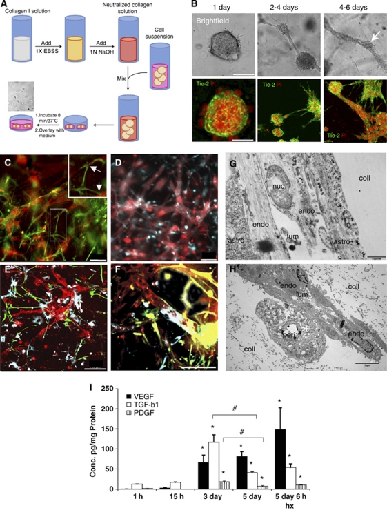
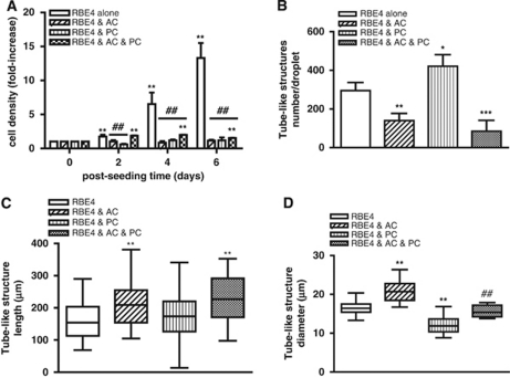
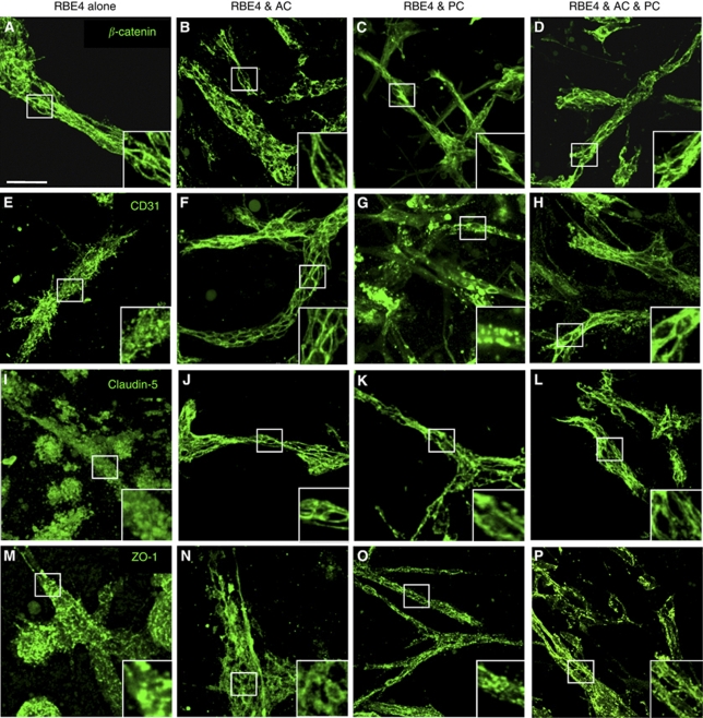
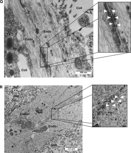
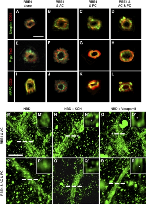
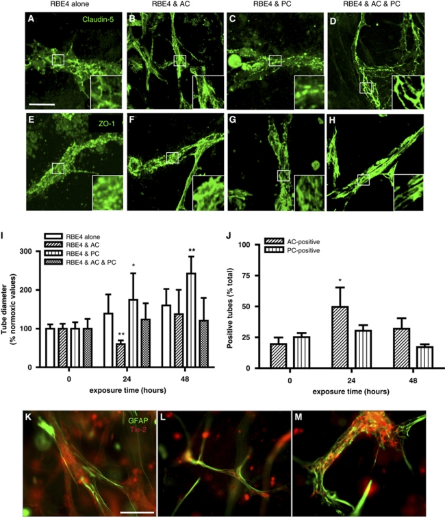
Understanding regulation of blood-brain barrier (BBB) is crucial to reduce/prevent its disruption during injury. As high brain complexity makes interpretation of in vivo data challenging, BBB studies are frequently performed using simplified in vitro models. However, many models fail to address the three-dimensional (3D) cellular interactions that occur in vivo, an important feature that may explain discrepancies in translation of in vitro data to the in vivo situation. We have designed and characterized an innovative 3D model that reproduces morphological and functional characteristics of the BBB in vivo and used it to investigate cellular interactions and contribution of astrocytes and pericytes to BBB development. Our model shows that both astrocytes and pericytes significantly suppress endothelial proliferation. In contrast, differential effects on tubulogenesis were observed with astrocytes reducing the number of tubes formed but increasing diameters and length, whereas pericytes had the opposite effect. Pericytes also induce proper localization of barrier proteins, lumen polarization, and functional activity of ATP-binding cassette (ABC) transporters similar to astrocytes, but the presence of both cells is required to maintain optimal barrier characteristics during hypoxic exposure. This model is simple, dynamic, and convenient to study many aspects of BBB function and represents an exciting new tool to address open questions of BBB regulation.


Figures

Figure 1
Figure 1
Collagen three-dimensional (3D) matrix supports tube formation, appropriate cellular interactions, and time-dependent expression of angiogenic molecules. (A) Diagrammatical scheme of the experimental protocol used for the collagen-based 3D cell culture. (B) Within the collagen matrix, RBE4 cells undergo morphogenesis first forming cyst-like structures, then connections between the cysts that finally result in a complex network of multicellular tube-like structures. The RBE4 cells were stained with the cell-specific marker Tie-2 (green), and cell nuclei were stained with propidium iodide (PI, red). Scale bar=50 μm. (C, D) Astrocytes and pericytes interact with RBE4 tubes in a similar manner as the in vivo situation. Note the glial fibrillary acidic protein (GFAP)-positive processes formed by astrocytes (green) over the RBE4 tube-like structures (red). Insert shows magnified image with arrows to highlight this interaction. In contrast, pericytes (DiD stain in blue) overlap RBE4 tubes. Scale bar=100 μm. Low (E) and high power images (F) show that the presence of both astrocytes and pericytes did not alter tube morphology or cellular interactions. Astrocyte processes (green) and pericyte overlap (blue) are easily visible at the vessel walls. Scale bar=100 μm. (G, H) Electron micrograph pictures of tubes within the matrix (coll) show the close physical contacts between RBE4 tubes (endo), astrocytes (astro, G), and pericytes (H, peri) as well as the presence of a lumen (lum). (I) Expression profiles of select angiogenic molecules show that secreted levels of VEGF, TGF-β1, and PDGF are significantly induced after 3 days of culture. Hypoxia further induces VEGF secretion, whereas TGF-β1 and PDGF concentrations remain unaffected. n=3; *P<0.05 compared with 1 hour time point, #P<0.05 at 3 days compared with 5-day time point.
Figure 2
Figure 2
Astrocytes (AC) and pericytes (PC) differentially modulate RBE4 tube morphogenesis. (A) Cell density continuously increased in RBE4 monocultures. The AC or PC cocultures inhibited the increase in cell density compared with RBE4 monocultures, whereas the presence of both cells (black and white squares bars) supported a modest increase after 2 days of culture. n=4; **P<0.01 compared with 0 day time point; ##P<0.01 compared with RBE4 monocultures. (B) AC and PC differentially modulate the number of RBE4 tubes formed after 6 days. Note the decreased number of tubes in the AC and three-cell cocultures compared with the PC group. n=4 ; *P<0.05; **P<0.01 and ***P<0.001 compared with RBE4 cells alone. (C) AC and three-cell cocultures induce longer tube-like structures. n=30, **P<0.01 compared with RBE4 cell group. (D) The AC induces larger diameter tubes, and PC induces thinner diameter tubes. The presence of both AC and PC resulted in similar diameter size as RBE4 monocultures. n=30; **P<0.01 compared with RBE4 monocultures, ##P<0.01 compared with two-cell cocultures.
Figure 3
Figure 3
Astrocytes (AC) and pericytes (PC) induce barrier maturation phenotype. β-Catenin (AD), CD31 (EH), claudin-5 (IL), and zonula occludens-1 (ZO-1) (MP) immunocytochemistry fluorescent micrograph pictures obtained from RBE4 cells cultured alone or in the presence of AC and PC. Note the absence of distinct tight junctions (TJs) localization at cell–cell borders in RBE4 monocultures as seen with both claudin-5 (I) and ZO-1 (M). Inserts are magnified images of regions highlighted by boxed areas. Scale bar=50 μm. Electron micrograph pictures from 5 day cocultures (Q) and RBE4 monocultures (R). Boxed out regions are shown as magnified pictures on the right-hand side. In the coculture image (Q), note the presence of electron-dense particles at the cell border (arrowheads), illustrating the presence of a TJ structure. Scale bar=500 nm. In contrast, note the absence of such particles at cell–cell borders in monocultures (R), indicating the absence of TJ structures in endothelial cells cultured alone. Scale bar=2 μm. Endo, endothelial cells; Coll, collagen matrix; Ap, astrocyte/pericyte.
Figure 3
Figure 3
Astrocytes (AC) and pericytes (PC) induce barrier maturation phenotype. β-Catenin (AD), CD31 (EH), claudin-5 (IL), and zonula occludens-1 (ZO-1) (MP) immunocytochemistry fluorescent micrograph pictures obtained from RBE4 cells cultured alone or in the presence of AC and PC. Note the absence of distinct tight junctions (TJs) localization at cell–cell borders in RBE4 monocultures as seen with both claudin-5 (I) and ZO-1 (M). Inserts are magnified images of regions highlighted by boxed areas. Scale bar=50 μm. Electron micrograph pictures from 5 day cocultures (Q) and RBE4 monocultures (R). Boxed out regions are shown as magnified pictures on the right-hand side. In the coculture image (Q), note the presence of electron-dense particles at the cell border (arrowheads), illustrating the presence of a TJ structure. Scale bar=500 nm. In contrast, note the absence of such particles at cell–cell borders in monocultures (R), indicating the absence of TJ structures in endothelial cells cultured alone. Scale bar=2 μm. Endo, endothelial cells; Coll, collagen matrix; Ap, astrocyte/pericyte.
Figure 4
Figure 4
Astrocytes and pericytes induce tube polarity and P-glycoprotein (P-gp) activity. (AL) Immunocytochemistry double staining and subsequent z-stack image reconstruction was used to obtain cross-sections of tube-like structures formed in astrocyte and pericyte cocultures. CD31 and Tie-2, endothelial markers that are not polarized, enable us to view the endothelial cells. Utrophin localization (AD) occurred on the basolateral side of the tubes in all groups, whereas the presence of astrocytes and pericytes were required to induce proper luminal polarity, as observed with P-gp (EH) and multidrug resistant protein-2 (MRP2) (IL) staining. Scale bar=10 μm. (MR) Live imaging combined with z-stack image reconstruction was used to obtain fluorescent micrograph pictures of coculture tube cross-sections after incubation with (N-(4-nitrobenzofurazan-7-yl)--Lys8)-cyclosporin A (NBD-CsA), a substrate for P-gp, in absence (M, P) or the presence of the P-gp inhibitors KCN (N, Q) or verapamil (O, R). Note the absence of NBD-CsA accumulation within the lumen (indicated with an asterisk) in the presence of either KCN or verapamil in high power images (N', O', Q', and R'). Scale bar=50 μm.
Figure 5
Figure 5
Astrocytes (AC) and pericytes (PC) differentially modulate RBE4 tube responses to hypoxic stimulus. (AH) The presence of both AC and PC is required to maintain tight junctions (TJs) complex localization at cell–cell borders in cultures exposed to prolonged hypoxia (1% O2 for 48 hours). Note that astrocyte cocultures (AC) appear to support better maintenance than pericyte cocultures (PC), as seen in high power inserts. Scale bar=50 μm. (I) The AC and PC modulate tube diameter in hypoxic cultures. Diameters are expressed as the percentage of their respective normoxic values. Note the transient decrease in diameter observed in AC cocultures, whereas PC cocultures induce a continuous increase in diameter over time. The pericyte effect is blunted in the three-cell cocultures. n=20; *P<0.05 and **P<0.01 against normoxic values. (J) Hypoxia transiently increases AC coverage of endothelial tubes, whereas no significant alteration in PC coverage was observed. *P<0.05 compared with normoxic values, n=5. (KM) Hypoxia acts as a biphasic stimulus for astrocyte–endothelial interactions. Immunocytochemistry confirms increased glial fibrillary acidic protein (GFAP+) staining (green) over the RBE4 tubes (red) after 24 hours (L) compared with normoxia (K). Prolonged exposure (48 hours) decreases GFAP+ staining over the tubes and induces an increase in tube diameter. Scale bar=50 μm.

References

    1. Al Ahmad A, Gassmann M, Ogunshola OO. Maintaining blood-brain barrier integrity: pericytes perform better than astrocytes during prolonged oxygen deprivation. J Cell Physiol. 2009;218:612–622. - PubMed
    1. Baldessari D, Mione M. How to create the vascular tree? (Latest) help from the zebrafish. Pharmacol Ther. 2008;118:206–230. - PubMed
    1. Banks WA, Ercal N, Price TO. The blood-brain barrier in neuroAIDS. Curr HIV Res. 2006;4:259–266. - PubMed
    1. Bauer B, Hartz AM, Fricker G, Miller DS. Pregnane X receptor up-regulation of P-glycoprotein expression and transport function at the blood-brain barrier. Mol Pharmacol. 2004;66:413–419. - PubMed
    1. Bell E, Ivarsson B, Merrill C. Production of a tissue-like structure by contraction of collagen lattices by human fibroblasts of different proliferative potential in vitro. Proc Natl Acad Sci USA. 1979;76:1274–1278. - PMC - PubMed

Publication types

MeSH terms

Substances