. 2010 May 1;129(2):99-109.
doi: 10.1007/s00435-010-0103-y.

The catecholaminergic nerve plexus of Holothuroidea

Affiliations

The catecholaminergic nerve plexus of Holothuroidea

Carlos A Díaz-Balzac et al. Zoomorphology. .

Abstract

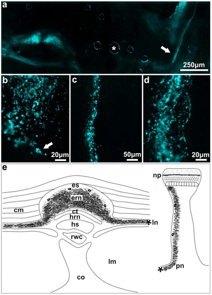
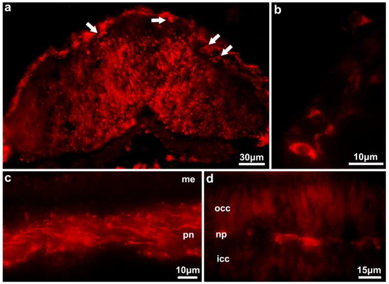
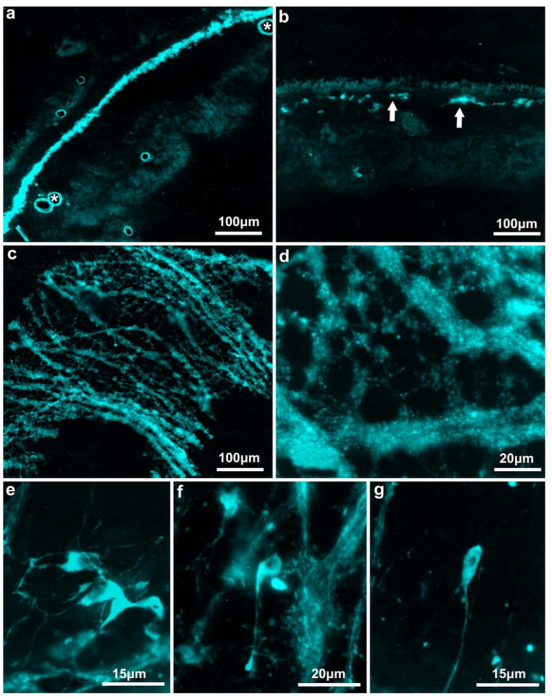
Catecholamines have been extensively reported to be present in most animal groups, including members of Echinodermata. In this study, we investigated the presence and distribution of catecholaminergic nerves in two members of the Holothuroidea, Holothuria glaberrima (Selenka, 1867) (Aspidochirotida, Holothuroidea) and Holothuria mexicana (Ludwig, 1875) (Aspidochirotida, Holothuroidea), by using induced fluorescence for catecholamines on tissue sections and immunohistochemistry with an antibody that recognizes tyrosine hydroxylase. The presence of a catecholaminergic nerve plexus similar in distribution and extension to those previously reported in other members of Echinodermata was observed. This plexus, composed of cells and fibers, is found in the ectoneural component of the echinoderm nervous system and is continuous with the circumoral nerve ring and the radial nerves, tentacular nerves, and esophageal plexus. In addition, fluorescent nerves in the tube feet are continuous with the catecholaminergic components of the radial nerve cords. This is the first comprehensive report on the presence and distribution of catecholamines in the nervous system of Holothuroidea. The continuity and distribution of the catecholaminergic plexus strengthen the notion that the catecholaminergic cells are interneurons, since these do not form part of the known sensory or motor circuits and the fluorescence is confined to organized nervous tissue.


Figures

Fig. 1
Fig. 1
Holothuria glaberrima (Holothuroidea). Catecholamine-induced fluorescence in the circumoral nerve ring. a Transverse section at the level of the circumoral nerve ring showing the network of catecholaminergic fibers. b Catecholaminergic cells (arrow) present within the fiber network, which are better distinguishable in its border
Fig. 2
Fig. 2
Holothuria glaberrima (Holothuroidea). Catecholamine-induced fluorescence in the tentacles. a, b Transverse section through the tentacle showing catecholamine-in-duced fluorescence in the buccal nerve (arrow) and in the buccal cylindrical fenestrated sheath (arrowheads). c Diagram depicting the distribution of the catecholaminergic plexus within the tentacle of Holothuroidea. bfs buccal cylindrical fenestrated sheath, bn buccal nerve, cct central connective tissue, ect external connective tissue, ict inner connective tissue, lu lumen, me mesothelium
Fig. 3
Fig. 3
Holothuria glaberrima (Holothuroidea). Catecholamine-induced fluorescence in the radial nerve cord and tube feet. a Transverse section of the body wall showing the induced fluorescence present in the ectoneural portion of the radial nerve cord and in the podial nerve (arrow). No fluorescence was present in the hyponeural component of the radial nerve cord. The circular structures (*) are artifacts produced by air bubbles introduced during the SPG procedure. b A section of the radial nerve at higher magnification showing details of the fiber network and a cell (arrow) present in the ectoneural component. c The podial nerve shows catecholamine-induced fluorescence, as can be observed in a longitudinal section. d A higher magnification of individual fluorescent fibers detected in the podial nerve. e Diagram showing the catecholaminergic containing structures within the radial nerve cord and tube feet. The anatomical continuity between the two nerves is also depicted as lateral nerves, branches from the radial nerve (*), which form the podial nerve as it enters the tube feet. cm circular muscle, co coelom, ct connective tissue, ern ectoneural component of the radial nerve, es epineural sinus, hrn hyponeural component of the radial nerve, hs hyponeural sinus, lm longitudinal muscle, ln lateral nerve, np nerve plate, pn podial nerve, rwc radial water vascular canal
Fig. 4
Fig. 4
Holothuria glaberrima (Holothuroidea). Antityrosine hydroxylase immunoreactivity distribution within the radial nerve cord and tube feet. a Transverse section through the body wall showing anti-TH immunoreactivity in Wbers and cells (arrows) of the ectoneural component of the radial nerve. b Anti-TH immunoreactive cells in the ectoneural component of the radial nerve. c Anti-TH immunopositive Wbers present within the podial nerve. d Anti-TH immunopositive Wbers located in the nerve plate of the tube feet's disk. icc inner cell cluter, me mesothelium, np nerve plate, pn podial nerve, occ outer cell cluster
Fig. 5
Fig. 5
Holothuria glaberrima (Holothuroidea). Catecholamineinduced fluorescence in the esophagus. The network of fluorescent fibers forming the catecholaminergic esophageal plexus is shown in transverse sections (a) at the level of the esophagus as a continuous layer of fluorescence observed between the muscular and submucosal layers, and (b) at the level of the esophagus-small intestine connection as discontinuous bundles (arrows) that will eventually disappear at a more posterior location. c Tangential sections of the esophagus showing the catecholaminergic fiber bundles forming the plexus. d Higher magnification of the catecholaminergic fiber tract. e-g Examples of the different morphologies exhibited by cells of the esophageal plexus. The circular structures (*) are artifacts produced by air bubbles introduced during the SPG procedure
Fig. 6
Fig. 6
Diagram depicting the catecholaminergic plexus in the Holothuroidea. The plexi in the circumoral nerve ring (nr), ectoneural component of the radial nerve (ern), buccal nerve (bn), podial nerve (pn), and esophagus (op) are continuous with each other, forming the structure shown in the diagram. An emphasis is done in the continuity and extension of the catecholaminergic plexus, and because of simplification we have omitted some details (such as ramification of the buccal nerve or the gradual disappearance of the esophageal plexus)

References

    1. Bishop CD, Burke RD. Ontogeny of the holothurian larval nervous system: evolution of larval forms. Dev Genes Evol. 2007;217(8):585–592. - PubMed
    1. Bouland C, Massin C, Jangoux M. The fine structure of the buccal tentacles of Holothuria forskali (Echinodermata, Holothuroidea) Zoomorphology. 1982;101:133–149.
    1. Bradford HF. Chemical neurobiology: an introduction to neurochemistry. Freeman; New York: 1986.
    1. Buznikov GA, Peterson RE, Nikitina LA, Bezuglov VV, Lauder JM. The pre-nervous serotonergic system of developing sea urchin embryos and larvae: pharmacologic and immunocytochemical evidence. Neurochem Res. 2005;30(6–7):825–837. - PubMed
    1. Cobb JLS. The distribution of mono-amines in the nervous system of echinoderms. Comp Biochem Physiol. 1969a;28:967–971.

LinkOut - more resources