

Ameloblastic fibro-odontoma: a diagnostic challenge
- PMID: 20827417
- PMCID: PMC2933904
- DOI: 10.1155/2010/104630
Ameloblastic fibro-odontoma: a diagnostic challenge
Abstract
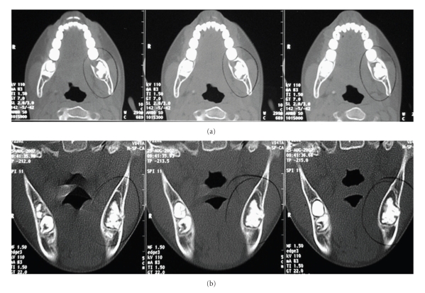
An 11-year-old girl presented to our department to have a second opinion regarding a lesion involving her left mandible. She had previously undergone several radiographic exams including panoramic, helical, and cone-beam computed tomography. Radiographic examinations revealed a well-defined radiolucent region, which contained an irregular radiopaque mass of 3 cm in diameter, localized to the left angle of the mandible. Our presumptive diagnosis was complex odontoma. Excisional biopsy was performed, and microscopic features showed strands and islands of odontogenic epithelium showing peripheral palisading and loosely arranged central cells, identical to stellate reticulum, embedded in a myxoid cell-rich stroma resembling the dental papilla. Dentin and enamel were also presented. The diagnosis was ameloblastic fibro-odontoma, which is a rare mixed odontogenic tumor, derived from epithelial and ectomesenchymal elements that form the dental tissues.
Figures


Similar articles
-
Extensive Mandibular Ameloblastic Fibro-Odontoma.J Craniofac Surg. 2016 Sep;27(6):e563-5. doi: 10.1097/SCS.0000000000002869. J Craniofac Surg. 2016. PMID: 27428924
-
Ameloblastic Fibro-Odontoma.Fetal Pediatr Pathol. 2023 Apr;42(2):281-284. doi: 10.1080/15513815.2022.2088910. Epub 2022 Jun 24. Fetal Pediatr Pathol. 2023. PMID: 35748698
-
Ameloblastic fibro-odontoma with chondroid tissue formation.Contemp Oncol (Pozn). 2018;22(1):50-53. doi: 10.5114/wo.2018.74395. Epub 2018 Apr 3. Contemp Oncol (Pozn). 2018. PMID: 29692665 Free PMC article.
-
Large ameloblastic fibro-odontoma in an 18-year-old girl and review of literature.BMJ Case Rep. 2012 Nov 19;2012:bcr-2012-007160. doi: 10.1136/bcr-2012-007160. BMJ Case Rep. 2012. PMID: 23166172 Free PMC article. Review.
-
[The ameloblastic fibro-odontoma].Dtsch Zahnarztl Z. 1991 Jan;46(1):71-3. Dtsch Zahnarztl Z. 1991. PMID: 1811980 Review. German.
Cited by
-
Aggressive ameloblastic fibro-odontoma assessment with CBCT and treatment.Eur Arch Paediatr Dent. 2013 Jun;14(3):179-84. doi: 10.1007/s40368-013-0032-9. Epub 2013 Apr 30. Eur Arch Paediatr Dent. 2013. PMID: 23633233
-
Ameloblastic fibro-odontoma.BMJ Case Rep. 2015 Jun 4;2015:bcr2015209739. doi: 10.1136/bcr-2015-209739. BMJ Case Rep. 2015. PMID: 26045519 Free PMC article.
-
Anomalous morphology of an ectopic tooth in the maxillary sinus on three-dimensional computed tomography images.J Radiol Case Rep. 2013 Feb 1;7(2):11-6. doi: 10.3941/jrcr.v7i2.1227. Print 2013 Feb. J Radiol Case Rep. 2013. PMID: 23705035 Free PMC article.
-
Ameloblastic Fibro-odontome (AFO) of the Mandible: A Case Report.J Clin Diagn Res. 2014 Jan;8(1):260-2. doi: 10.7860/JCDR/2014/5402.3942. Epub 2014 Jan 12. J Clin Diagn Res. 2014. PMID: 24596790 Free PMC article.
-
Clinical and radiological profile of ameloblastic fibro-odontoma: an update on an uncommon odontogenic tumor based on a critical analysis of 114 cases.Head Neck Pathol. 2013 Mar;7(1):54-63. doi: 10.1007/s12105-012-0397-9. Epub 2012 Sep 22. Head Neck Pathol. 2013. PMID: 23001451 Free PMC article.
References
-
- Hamner JE, III, Pizer ME. Ameloblastic odontoma. Report of two cases. American Journal of Diseases of Children. 1968;115(3):332–336. - PubMed
-
- Hawkins PL, Sadeghi EM. Ameloblastic fibro-odontoma: report of a case. Journal of Oral and Maxillofacial Surgery. 1986;44(12):1014–1019. - PubMed
-
- Wu PC, Chan KW. A survey of tumours of the jawbones in Hong Kong Chinese: 1963–1982. British Journal of Oral and Maxillofacial Surgery. 1985;23(2):92–102. - PubMed
-
- O’Brien FV. Ameloblastic odontome. A case report. British Dental Journal. 1971;131(2):71–72. - PubMed
-
- Okura M, Nakahara H, Matsuya T. Treatment of ameloblastic fibro-odontoma without removal of the associated impacted permanent tooth: report of cases. Journal of Oral and Maxillofacial Surgery. 1992;50(10):1094–1097. - PubMed
Publication types
LinkOut - more resources
Full Text Sources
