. 2010 Oct;40(10):2816-29.
doi: 10.1002/eji.201040384.

Murine model of chronic L. (Viannia) panamensis infection: role of IL-13 in disease

Affiliations

Murine model of chronic L. (Viannia) panamensis infection: role of IL-13 in disease

Tiago M Castilho et al. Eur J Immunol. 2010 Oct.

Abstract

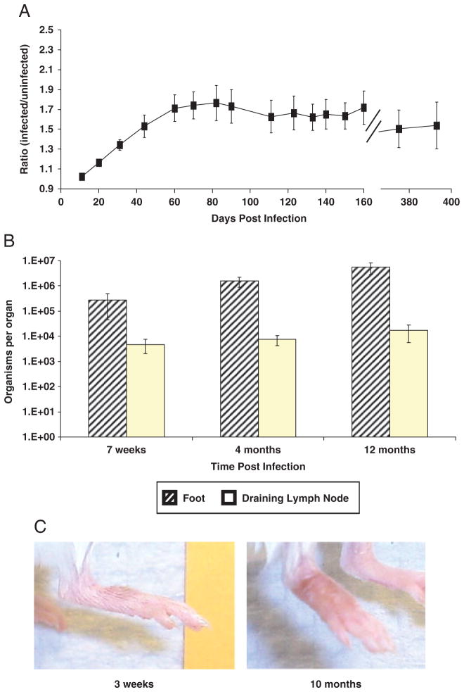
Leishmania (Viannia) organisms are the most prevalent etiologic agents of human cutaneous leishmaniasis in the Americas. Nevertheless, our knowledge of the immunological mechanisms exploited by L. (Viannia) organisms remains limited and the mechanisms underlying disease are not well understood. Here, we report the development of a BALB/c mouse model of L. (V.) panamensis infection that is able to reproduce chronic disease, with persistent infection and clinically evident lesions for over 1 year. The immune response of the mouse resembles that found for L. (V.) panamensis-infected patients with chronic and recurrent lesions, presenting a mixed Th1/Th2 response with the presence of TNF-α, IFN-γ, IL-10 and IL-13. Using immunodeficient mice, the critical role for IL-13 and/or IL-4Rα in determining susceptibility to chronic infection was evident. With the induction of healing in the immunodeficient mice, increases in IFN-γ and IL-17 were found, concomitant with parasite control and elimination. Specifically, increases in CD4(+) (but not CD8(+)) T cells producing IFN-γ were observed. These results suggest that IL-13 represents an important target for disease control of L. (V.) panamensis infection. This murine model should be useful to further understand the pathology associated with chronic disease and to develop methods for the treatment and prevention of leishmaniasis caused by L. (Viannia) parasites.


Conflict of interest statement

Conflict of interest: The authors declare no financial or commercial conflict of interest.

Figures

Figure 1
Figure 1
Stationary phase L. (V.) panamensis promastigotes generate chronic lesions in BALB/c mice. Mice were intradermally infected (5 × 10⁴ late log phase percoll-purified organisms) on the top of the right rear hind foot. Course of infection was monitored by measuring (A) ratio of lesion development in BALB/c footpad compared with the contralateral uninfected foot (n = 10), and by (B) parasite burden analyses of feet (diagonal-stripped bars) or pooled draining popliteal and inguinal LN (open bars, n = 4–5). (C) Representative median lesions of infection at 3 wk or 10 months after infection. Data show mean ± SEM and are representative of at least two independent experiments.
Figure 2
Figure 2
Human natural infection presents a clinical spectrum (from chronic and recurrent lesions to asymptomatic infection) characterized by an immune response constituted by a mixed pro- and anti-inflammatory cytokine profile. (A) Cytokine production by PBMC from patients with active chronic (CH) and recurrent disease (RE), asymptomatically infected individuals (AS) and healthy controls (HC). ANOVA (p < 0.001 for all cytokines) and Duncan’s test for multiple comparison, *p < 0.01. (B) Regression analyses of cytokine responses across the spectrum of disease caused by L. (V.) panamensis. Regression analyses of cytokine production were conducted only in infected individuals (chronic, recurrent and asymptomatic) using Pearson correlation analysis. p-Values of less than 0.05 are considered significant.
Figure 3
Figure 3
L. (V.) panamensis infection in BALB/c does not require B cells. (A) Lesion development was monitored by lesion development of infected footpad in BALB/c WT (squares) or B-cell-deficient, JhD (circles) mice, infected with 5 × 10⁴ stationary phase purified promastigotes, a minimum of ten mice of each group was analyzed for lesion development. Results are representative of three independent experiments. Parasite burden was determined at (B) 2 or (C) 4 months post-infection (n = 4/group/determination). Data show mean ± SEM, *p < 0.05 by unpaired Wilcoxon test.
Figure 4
Figure 4
L. (V.) panamensis infection in BALB/c requires IL-13 and IL-4Rα. Course of infection was monitored by (A) footpad lesion development in WT (squares), and in IL-4Rα (triangles) or IL-13- (open circles) deficient BALB/c mice (data show at least four mice per group and are representative of three independent experiments). *(IL-13 KO) and +(IL-4Rα KO) indicate p < 0.001 by unpaired Wilcoxon test compared with WT controls. Parasite burden was determined at (B) 25 days and (C) 60 days post-infection in BALB/c WT (filled bars), IL-13 KO (diagonally stripped bars) and IL-4Rα KO (open bars). The numbers of mice with positive parasite burden in each group are indicated beneath the bars. *p < 0.03 by unpaired Wilcoxon test and error bars represent standard error of the mean (n = 4). Similar results were observed at 180 and 360 days post-infection.
Figure 5
Figure 5
Response to infection with L. (V.) panamensis of WT and IL-13 or IL-4Rα-deficient mice. Cytokine responses of WT, IL-13- or IL-4Rα-deficient BALB/c mice at different times post-infection, as indicated. ELISA analyses for cytokines were performed on supernatants of spleen-derived lymphocytic cells stimulated with Con A or pLAg (as indicated in the Materials and methods section). Data show mean ± SEM. *(IL-13 KO) and +(IL-4Rα KO) indicate p < 0.05 by Student’s T-test. Similar results were obtained in another independent experiment at 3.5 wk. The IL-4 production by lymphocytes of IL-4Rα-deficient mice is not shown. However, consistent with the previous observations [68], IL-4 levels were found to be >300 pg/mL at all time points.
Figure 6
Figure 6
Lack of IL-13 signaling leads to increases in CD4+IFN-γ+ T cells. IFN-γ responses of WT or IL-13-deficient BALB/c mice at 3.5 wk post-infection. FACS analyses for IFN-γ were performed as indicated in the Materials and methods section (gating strategy is shown in Supporting Information Fig. 4) using spleen lymphocytes stimulated with pLAg. Lymphocyte populations (based on the side and forward scatter) were sorted for either surface (A) CD4 or (B) CD8 expression (as indicated) and examined for IFN-γ synthesis. Results are representative of two independent experiments (n = 4 mice/group). (C) Ratio of % IFN-γ+ cells divided by % IFN-γ+ cells in the WT group is shown (average of points from multiple experiments), error bars indicate SD, and *p < 0.03 by Wilcoxon test.

References

    1. Murray HW, Flanders KC, Donaldson DD, Sypek JP, Gotwals PJ, Liu J, Ma X. Antagonizing deactivating cytokines to enhance host defense and chemotherapy in experimental visceral leishmaniasis. Infect Immun. 2005;73:3903–3911. - PMC - PubMed
    1. WHO. Magnitude of the Problem. 2009 http://www.who.int/leishmaniasis/burden/magnitude/burden_magnitude/en/in....
    1. Grimaldi G, Jr, Tesh RB, McMahon-Pratt D. A review of the geographic distribution and epidemiology of leishmaniasis in the New World. Am J Trop Med Hyg. 1989;41:687–725. - PubMed
    1. Pirmez C, Yamamura M, Uyemura K, Paes-Oliveira M, Conceicao-Silva F, Modlin RL. Cytokine patterns in the pathogenesis of human leishmaniasis. J Clin Invest. 1993;91:1390–1395. - PMC - PubMed
    1. Bosque F, Saravia NG, Valderrama L, Milon G. Distinct innate and acquired immune responses to Leishmania in putative susceptible and resistant human populations endemically exposed to L. (Viannia) panamensis infection. Scand J Immunol. 2000;51:533–541. - PubMed

Publication types

MeSH terms