

Distribution and quantitative changes in amounts of aquaporin 1, 5 and 9 in the pig uterus during the estrous cycle and early pregnancy
- PMID: 20828411
- PMCID: PMC2944173
- DOI: 10.1186/1477-7827-8-109
Distribution and quantitative changes in amounts of aquaporin 1, 5 and 9 in the pig uterus during the estrous cycle and early pregnancy
Abstract
Background: Aquaporins (AQPs) are a family of membrane channel proteins that facilitate bulk water transport. To date, 11 isoforms of AQPs have been reported to be expressed in the female and male reproductive systems. The purpose of our study was to determine the localization and quantitative changes in the expression of AQP1, 5 and 9 within the pig uterus during different stages of the estrous cycle and early pregnancy.
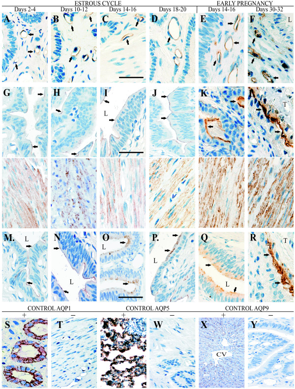
Methods: Immunoperoxidase and semi-quantitative immunoblotting techniques were used to examine the distribution and changes in amounts of AQP1, AQP5 and AQP9 in uteral cells of pigs at the early (Days 2-4), middle (10-12), late (14-16) stage of the luteal phase and late (18-20) stage of the follicular phase of the estrous cycle as well as on Days 14-16 and 30-32 of gestation (the onset and the end of implantation process).
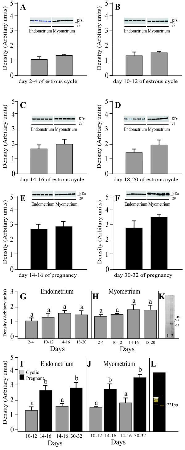
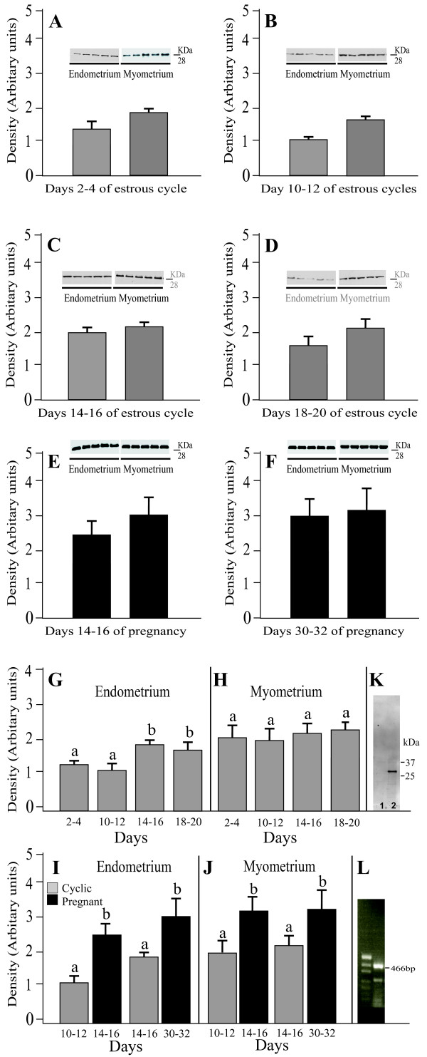
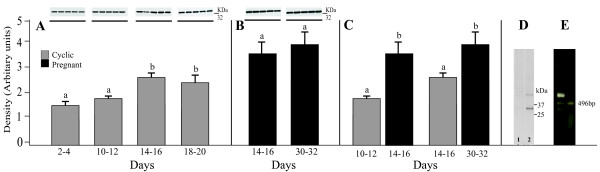
Results: The results demonstrated that AQP1, 5, and 9 were clearly detected in all studied stages of the estrous cycle and pregnancy. AQP1 was localized within uterine blood vessels. In cyclic gilts, endometrial and myometrial expression of AQP1 protein did not change significantly but increased during gestation. AQP5 was localized in smooth muscle cells and uterine epithelial cells. Endometrial expression of AQP5 protein did not change significantly between Days 2-4 and 10-12 of the estrous cycle but increased on Days 14-16 and 18-20 as well as during early pregnancy. Myometrial expression of AQP5 did not differ significantly during the estrous cycle but increased in the pregnancy. The anti-AQP9 antibody labeled uterine epithelial cells of uterus. Endometrial expression of AQP9 did not change significantly between Days 2-4 and 10-12 of the estrous cycle but increased on Days 14-16 and 18-20 as well as during early pregnancy.
Conclusions: The results suggest that a functional and distinctive collaboration exists among diverse AQPs in water handling during the different uterine phases in the estrous cycle and early pregnancy.
Figures
Similar articles
-
Progesterone, estradiol, arachidonic acid, oxytocin, forskolin and cAMP influence on aquaporin 1 and 5 expression in porcine uterine explants during the mid-luteal phase of the estrous cycle and luteolysis: an in vitro study.Reprod Biol Endocrinol. 2015 Feb 18;13:7. doi: 10.1186/s12958-015-0004-5. Reprod Biol Endocrinol. 2015. PMID: 25884220 Free PMC article.
-
Fluctuation of aquaporin 1, 5, and 9 expression in the pig oviduct during the estrous cycle and early pregnancy.J Histochem Cytochem. 2011 Apr;59(4):419-27. doi: 10.1369/0022155411400874. J Histochem Cytochem. 2011. PMID: 21411812 Free PMC article.
-
A collaboration of aquaporins handles water transport in relation to the estrous cycle in the bitch uterus.Theriogenology. 2009 Aug;72(3):310-21. doi: 10.1016/j.theriogenology.2009.01.023. Epub 2009 Apr 23. Theriogenology. 2009. PMID: 19395011
-
Uterine blood supply as a main factor involved in the regulation of the estrous cycle--a new theory.Reprod Biol. 2002 Jul;2(2):93-114. Reprod Biol. 2002. PMID: 14666152 Review.
-
The Expanding Role of Aquaporin-1, Aquaporin-3 and Aquaporin-5 as Transceptors: Involvement in Cancer Development and Potential Druggability.Int J Mol Sci. 2025 Feb 4;26(3):1330. doi: 10.3390/ijms26031330. Int J Mol Sci. 2025. PMID: 39941100 Free PMC article. Review.
Cited by
-
Testosterone Induces Increase in Aquaporin (AQP)-1, 5, and 7 Expressions in the Uteri of Ovariectomized Rats.J Membr Biol. 2015 Dec;248(6):1097-105. doi: 10.1007/s00232-015-9823-8. Epub 2015 Jul 22. J Membr Biol. 2015. PMID: 26198330
-
The Relevance of Aquaporins for the Physiology, Pathology, and Aging of the Female Reproductive System in Mammals.Cells. 2020 Dec 1;9(12):2570. doi: 10.3390/cells9122570. Cells. 2020. PMID: 33271827 Free PMC article. Review.
-
Progesterone, estradiol, arachidonic acid, oxytocin, forskolin and cAMP influence on aquaporin 1 and 5 expression in porcine uterine explants during the mid-luteal phase of the estrous cycle and luteolysis: an in vitro study.Reprod Biol Endocrinol. 2015 Feb 18;13:7. doi: 10.1186/s12958-015-0004-5. Reprod Biol Endocrinol. 2015. PMID: 25884220 Free PMC article.
-
Fluctuation of aquaporin 1, 5, and 9 expression in the pig oviduct during the estrous cycle and early pregnancy.J Histochem Cytochem. 2011 Apr;59(4):419-27. doi: 10.1369/0022155411400874. J Histochem Cytochem. 2011. PMID: 21411812 Free PMC article.
-
Reduced expression of aquaporin 9 in tubal ectopic pregnancy.J Mol Histol. 2013 Apr;44(2):167-73. doi: 10.1007/s10735-012-9471-6. Epub 2012 Dec 14. J Mol Histol. 2013. PMID: 23238960
References
-
- Verkman AS, Mitra AK. Structure and function of aquaporin water channels. Am J Physiol Renal Physiol. 2000;278:13–28. - PubMed
Publication types
MeSH terms
Substances
LinkOut - more resources
Full Text Sources
Molecular Biology Databases
