. 2010;2(6):576-86.
doi: 10.1159/000319909. Epub 2010 Sep 10.

Nuclease expression by Staphylococcus aureus facilitates escape from neutrophil extracellular traps

Affiliations

Nuclease expression by Staphylococcus aureus facilitates escape from neutrophil extracellular traps

Evelien T M Berends et al. J Innate Immun. 2010.

Abstract

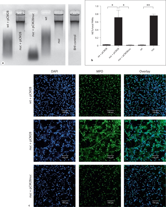
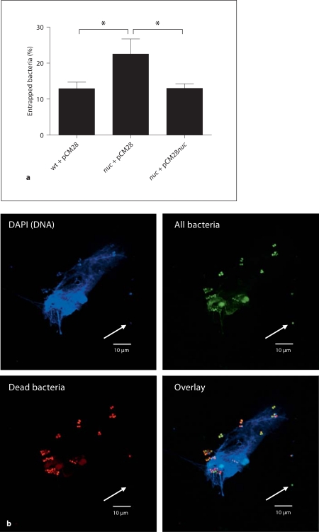
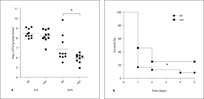
Neutrophils are key effectors of the host innate immune response against bacterial infection. Staphylococcus aureus is a preeminent human pathogen, with an ability to produce systemic infections even in previously healthy individuals, thereby reflecting a resistance to effective neutrophil clearance. The recent discovery of neutrophil extracellular traps (NETs) has opened a novel dimension in our understanding of how these specialized leukocytes kill pathogens. NETs consist of a nuclear DNA backbone associated with antimicrobial peptides, histones and proteases that provide a matrix to entrap and kill various microbes. Here, we used targeted mutagenesis to examine a potential role of S. aureus nuclease in NET degradation and virulence in a murine respiratory tract infection model. In vitro assays using fluorescence microscopy showed the isogenic nuclease-deficient (nuc-deficient) mutant to be significantly impaired in its ability to degrade NETs compared with the wild-type parent strain USA 300 LAC. Consequently, the nuc-deficient mutant strain was significantly more susceptible to extracellular killing by activated neutrophils. Moreover, S. aureus nuclease production was associated with delayed bacterial clearance in the lung and increased mortality after intranasal infection. In conclusion, this study shows that S. aureus nuclease promotes resistance against NET-mediated antimicrobial activity of neutrophils and contributes to disease pathogenesis in vivo.


Figures

Fig. 1
Fig. 1
S. aureus nuclease degrades calf thymus DNA and NETs. a Representative agarose gel of bacterial culture supernatants after incubation with calf thymus DNA to detect nuclease activity. Samples with nuclease activity show a smear of degraded DNA on the gel as is seen in the lanes representing the S. aureus LAC wild-type empty vector control (wt + pCM28), complemented nuc-mutant strain (nuc + pCM28nuc) and wild-type (no vector, wt). In the lanes with samples from nuc-mutant empty vector control (nuc + pCM28), nuc-mutant (no vector, nuc) and sterile BHI medium (negative control), the DNA is not degraded, indicating that no nuclease activity is present. b Quantification of NETs after coincubation of PMA-stimulated neutrophils with S. aureus LAC wild-type empty vector control (wt + pCM28), nuc-mutant empty vector control (nuc + pCM28) or complemented mutant strain (nuc + pCM28nuc), wild-type (no vector, wt) or nuc-mutant (no vector, nuc) at a MOI of 2 for 90 min. After coincubation, the slides were fixed and stained for MPO to visualize the NETs and mounted in DAPI to stain DNA. The amount of neutrophils that release NETs was counted per field of view and compared with the total amount of neutrophils. The results of 4 (wt + pCM28, nuc + pCM28, nuc + pCM28nuc) or 3 (wt, nuc) independent experiments were analyzed using a paired, one-tailed Student's t test. ∗ p < 0.05; ∗∗ p < 0.01. c Representative immunofluorescent micrograph of PMA-stimulated neutrophils coincubated with S. aureus LAC wild-type empty vector control (wt + pCM28), nuc-mutant empty vector control (nuc + pCM28) or complemented mutant strain (nuc + pCM28nuc). NETs were visualized with a primary rabbit-anti-MPO antibody and a secondary Alexa 488-labeled goat-anti-rabbit antibody (green). DNA is stained with DAPI (blue).
Fig. 2
Fig. 2
S. aureus nuclease facilitates evasion from NET entrapment. a Quantitative analysis of bacterial entrapment by activated neutrophils. FITC-labeled bacteria [S. aureus LAC wild-type empty vector control (wt + pCM28), nuc-mutant empty vector control (nuc + pCM28) or complemented mutant strain (nuc + pCM28nuc)] were coincubated with PMA-stimulated neutrophils at a MOI of 200 for 90 min at 37°C in 5% CO2. After incubation, the plates were centrifuged and the wells were carefully washed twice with fresh medium to remove bacteria that were not entrapped within the NETs. The percentage of entrapped bacteria was calculated as (A458/538 nm of wells containing neutrophils)/(A458/538 of wells without neutrophils) × 100. The results of 5 independent experiments were analyzed using a paired, one-tailed Student's t test. ∗ p < 0.05. b Representative fluorescent micrograph showing viability of S. aureus LAC nuc-mutant (nuc + pCM28) entrapped by or in close proximity to NETs. Live/Dead Bac-Light™ Bacterial Viability Kit (Invitrogen) was used to determine the viability of bacteria after coincubation with stimulated neutrophils. Similar bacterial killing within remaining NETs has been detected in case of the wild-type strain (data not shown). The green dye (SYTO 9) generally labels all bacteria – bacteria with intact as well as damaged membranes. In contrast, the red dye (propidium iodide) penetrates only bacteria with damaged membranes, causing a reduction in the green (SYTO 9) fluorescence stain. Note that bacteria entrapped by or in close proximity to the NETs are dead (red + green) whereas bacteria that are further away from the NETs remain alive (green, white arrow).
Fig. 3
Fig. 3
S. aureus nuclease mediates resistance against extracellular killing by neutrophils. a Representative immunofluorescent micrograph of neutrophils coincubated with S. aureus LAC nuc + pCM28 in the presence or absence of 10 μg/ml cytochalasin D (to block phagocytosis) showing that NETs are produced in the presence of cytochalasin D. NETs were visualized with a primary rabbit-anti-MPO antibody and a secondary Alexa 488-labeled goatanti-rabbit antibody (green). DNA is stained with DAPI (blue). b Bacterial growth inhibition after coincubation of S. aureus LAC wild-type empty vector control (wt + pCM28), nuc-mutant empty vector control (nuc + pCM28) or complemented mutant strain (nuc + pCM28nuc) with PMA-stimulated neutrophils. Phagocytosis was blocked by treatment of the cells with 10 μg/ml cytochalasin D, 20 min prior to infection. Data are presented as percentage of surviving bacteria compared with the respective bacterial growth control (100%). The results of 4 independent experiments were analyzed using a paired, one-tailed Student's t test. ∗∗ p < 0.01.
Fig. 4
Fig. 4
Formation of NETs in S. aureus-infected lungs in vivo. Representative immunofluorescent micrograph showing the presence of NETs in the alveolar space of murine lung sections 24 h after intranasal infection with 2 × 10⁸ CFU of S. aureus LAC wild-type. In the right panel of S. aureus-infected lung tissue, a cell in the alveolar space is visible (white arrow), which releases a mixture of CRAMP and DNA-histone complexes (NETs) into the surrounding (alveolar space at the top of the DAPI-stained nucleus). Those NETs are absent in lungs from the PBS control mice. NETs were visualized using a triple-staining of DAPI to stain DNA (blue), monoclonal mouse anti-H2A-H2B-DNA complex antibody followed by an Alexa 488-goatanti-mouse antibody (green) and rabbitanti-CRAMP antibody followed by Alexa 568-goat-anti-rabbit antibody (red).
Fig. 5
Fig. 5
S. aureus nuclease expression mediates pathogenesis in vivo. a Bacterial load (CFU) in lung tissue of 10 mice (pooled from 2 individual experiments with 5 mice each) at 6 and 24 h after intranasal infection with 2 × 10⁸ CFU S. aureus LAC wild-type (wt) or nuc-mutant (nuc) strain. Differences between the 2 groups were analyzed by using a unpaired, one-tailed Student's t test (∗ p < 0.05). b Survival of female CD-1 mice (n = 24, pooled from 4 individual experiments with 6 mice each) after intranasal infection with 3 or 4 × 10⁸ CFU of S. aureus LAC wild-type (wt) or nuc-mutant (nuc) strain. Comparison of survival curves was performed with the Gehan-Breslow-Wilcoxon test (∗ p < 0.05).

References

    1. Ogston A. Micrococcus poisoning. J Anat Physiol. 1882;16:526–567. - PMC - PubMed
    1. Lowy FD. Staphylococcus aureus infections. N Engl J Med. 1998;339:520–532. - PubMed
    1. Chambers HF, DeLeo FR. Waves of resistance: Staphylococcus aureus in the antibiotic era. Nat Rev Microbiol. 2009;7:629–641. - PMC - PubMed
    1. Kobayashi SD, Braughton KR, Palazzolo-Ballance AM, Kennedy AD, Sampaio E, Kristosturyan E, Whitney AR, Sturdevant DE, Dorward DW, Holland SM, Kreiswirth BN, Musser JM, DeLeo FR. Rapid neutrophil destruction following phagocytosis of Staphylococcus aureus. J Innate Immun. 2010 E-pub ahead of print. - PMC - PubMed
    1. Bronner S, Monteil H, Prévots G. Regulation of virulence determinants in Staphylococcus aureus: complexity and applications. FEMS Microbiol Rev. 2004;28:183–200. - PubMed

Publication types

MeSH terms

Substances