

Oxidative damage and the prevention of age-related cataracts
- PMID: 20829639
- PMCID: PMC2952186
- DOI: 10.1159/000316481
Oxidative damage and the prevention of age-related cataracts
Abstract
Purpose: Cataracts are often considered to be an unavoidable consequence of aging. Oxidative damage is a major cause or consequence of cortical and nuclear cataracts, the most common types of age-related cataracts.
Methods: In this review, we consider the different risk factors, natural history and etiology of each of the 3 major types of age-related cataract, as well as the potential sources of oxidative injury to the lens and the mechanisms that protect against these insults. The evidence linking different oxidative stresses to the different types of cataracts is critically evaluated.
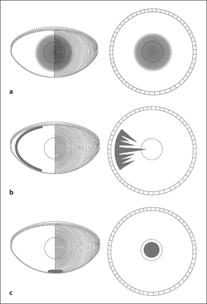
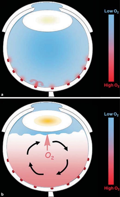
Results: We conclude from this analysis that the evidence for a causal role of oxidation is strong for nuclear, but substantially lower for cortical and posterior subcapsular cataracts. The preponderance of evidence suggests that exposure to increased levels of molecular oxygen accelerates the age-related opacification of the lens nucleus, leading to nuclear cataract. Factors in the eye that maintain low oxygen partial pressure around the lens are, therefore, important in protecting the lens from nuclear cataract.
Conclusions: Maintaining or restoring the low oxygen partial pressure around that lens should decrease or prevent nuclear cataracts.
Copyright 2010 S. Karger AG, Basel.
Figures
Similar articles
-
Etiology of posterior subcapsular cataracts based on a review of risk factors including aging, diabetes, and ionizing radiation.Int J Radiat Biol. 2020 Nov;96(11):1339-1361. doi: 10.1080/09553002.2020.1812759. Epub 2020 Sep 22. Int J Radiat Biol. 2020. PMID: 32897800 Review.
-
[Pathophysiology of cataract formation after vitrectomy].Klin Monbl Augenheilkd. 2010 Mar;227(3):175-80. doi: 10.1055/s-0029-1245271. Epub 2010 Mar 16. Klin Monbl Augenheilkd. 2010. PMID: 20234979 Review. German.
-
Reactive Oxygen Species and the Aging Eye: Specific Role of Metabolically Active Mitochondria in Maintaining Lens Function and in the Initiation of the Oxidation-Induced Maturity Onset Cataract--A Novel Platform of Mitochondria-Targeted Antioxidants With Broad Therapeutic Potential for Redox Regulation and Detoxification of Oxidants in Eye Diseases.Am J Ther. 2016 Jan-Feb;23(1):e98-117. doi: 10.1097/MJT.0b013e3181ea31ff. Am J Ther. 2016. PMID: 21048433 Review.
-
Lipid peroxidation and cataracts: N-acetylcarnosine as a therapeutic tool to manage age-related cataracts in human and in canine eyes.Drugs R D. 2004;5(3):125-39. doi: 10.2165/00126839-200405030-00001. Drugs R D. 2004. PMID: 15139774 Review.
-
Telomere Attrition in Human Lens Epithelial Cells Associated with Oxidative Stress Provide a New Therapeutic Target for the Treatment, Dissolving and Prevention of Cataract with N-Acetylcarnosine Lubricant Eye Drops. Kinetic, Pharmacological and Activity-Dependent Separation of Therapeutic Targeting: Transcorneal Penetration and Delivery of L-Carnosine in the Aqueous Humor and Hormone-Like Hypothalamic Antiaging Effects of the Instilled Ophthalmic Drug Through a Safe Eye Medication Technique.Recent Pat Drug Deliv Formul. 2016;10(2):82-129. doi: 10.2174/1872211309666150618104657. Recent Pat Drug Deliv Formul. 2016. PMID: 26084629 Review.
Cited by
-
Aging lens epithelium is susceptible to ferroptosis.Free Radic Biol Med. 2021 May 1;167:94-108. doi: 10.1016/j.freeradbiomed.2021.02.010. Epub 2021 Mar 17. Free Radic Biol Med. 2021. PMID: 33722625 Free PMC article.
-
Effects of senescent lens epithelial cells on the severity of age-related cortical cataract in humans: A case-control study.Medicine (Baltimore). 2016 Jun;95(25):e3869. doi: 10.1097/MD.0000000000003869. Medicine (Baltimore). 2016. PMID: 27336873 Free PMC article.
-
The Relationship Between Serum 25-Hydroxyvitamin D Levels and Nuclear Cataract in the Carotenoid Age-Related Eye Study (CAREDS), an Ancillary Study of the Women's Health Initiative.Invest Ophthalmol Vis Sci. 2015 Jul;56(8):4221-30. doi: 10.1167/iovs.15-16835. Invest Ophthalmol Vis Sci. 2015. PMID: 26132781 Free PMC article.
-
Switching to instant black coffee modulates sodium selenite-induced cataract in rats.Ger Med Sci. 2016 Apr 25;14:Doc05. doi: 10.3205/000232. eCollection 2016. Ger Med Sci. 2016. PMID: 27158251 Free PMC article.
-
Effect of vitreomacular adhesion and vitreous gel on age-related reduction of macular thickness: a retrospective observational study.BMJ Open. 2016 Sep 30;6(9):e012972. doi: 10.1136/bmjopen-2016-012972. BMJ Open. 2016. PMID: 27694490 Free PMC article.
References
-
- The Eye Diseases Prevalence Research Group Causes and prevalence of visual impairment among adults in the United States. Arch Ophthalmol. 2004;122:477–485. - PubMed
-
- West S. Epidemiology of cataract: accomplishments over 25 years and future directions. Ophthalmic Epidemiol. 2007;14:173–178. - PubMed
-
- Salm M, Belsky D, Sloan FA. Trends in cost of major eye diseases to Medicare, 1991 to 2000. Am J Ophthalmol. 2006;142:976–982. - PubMed
-
- Sasaki K, et al. Racial differences of lens transparency properties with aging and prevalence of age-related cataract applying a WHO classification system. Ophthalmic Res. 2004;36:332–340. - PubMed
Publication types
MeSH terms
Substances
Grants and funding
LinkOut - more resources
Full Text Sources
Other Literature Sources
Medical
