. 2010 Sep 7;5(9):e12580.
doi: 10.1371/journal.pone.0012580.

A mouse model of post-arthroplasty Staphylococcus aureus joint infection to evaluate in vivo the efficacy of antimicrobial implant coatings

Affiliations

A mouse model of post-arthroplasty Staphylococcus aureus joint infection to evaluate in vivo the efficacy of antimicrobial implant coatings

Nicholas M Bernthal et al. PLoS One. .

Abstract

Background: Post-arthroplasty infections represent a devastating complication of total joint replacement surgery, resulting in multiple reoperations, prolonged antibiotic use, extended disability and worse clinical outcomes. As the number of arthroplasties in the U.S. will exceed 3.8 million surgeries per year by 2030, the number of post-arthroplasty infections is projected to increase to over 266,000 infections annually. The treatment of these infections will exhaust healthcare resources and dramatically increase medical costs.

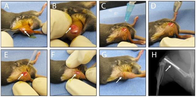
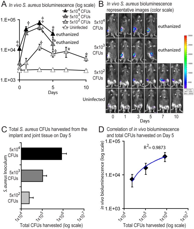
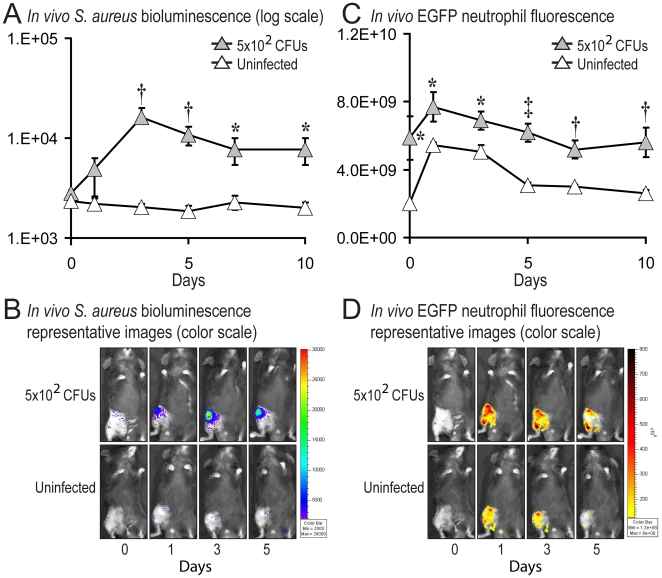
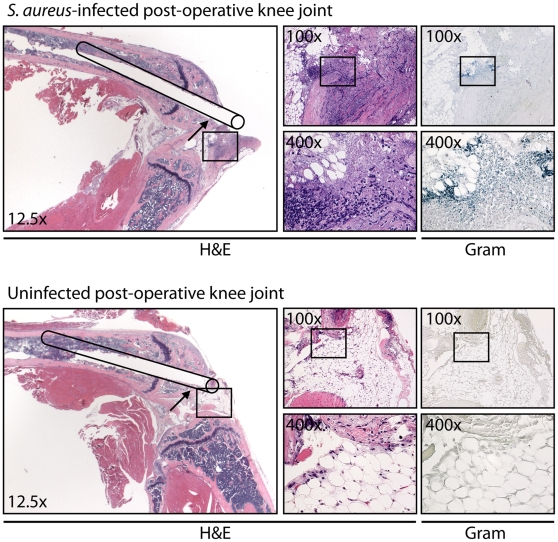
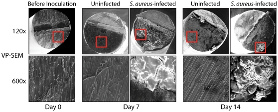
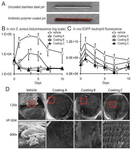
Methodology/principal findings: To evaluate novel preventative therapeutic strategies against post-arthroplasty infections, a mouse model was developed in which a bioluminescent Staphylococcus aureus strain was inoculated into a knee joint containing an orthopaedic implant and advanced in vivo imaging was used to measure the bacterial burden in real-time. Mice inoculated with 5x10(3) and 5x10(4) CFUs developed increased bacterial counts with marked swelling of the affected leg, consistent with an acute joint infection. In contrast, mice inoculated with 5x10(2) CFUs developed a low-grade infection, resembling a more chronic infection. Ex vivo bacterial counts highly correlated with in vivo bioluminescence signals and EGFP-neutrophil fluorescence of LysEGFP mice was used to measure the infection-induced inflammation. Furthermore, biofilm formation on the implants was visualized at 7 and 14 postoperative days by variable-pressure scanning electron microscopy (VP-SEM). Using this model, a minocycline/rifampin-impregnated bioresorbable polymer implant coating was effective in reducing the infection, decreasing inflammation and preventing biofilm formation.

Conclusions/significance: Taken together, this mouse model may represent an alternative pre-clinical screening tool to evaluate novel in vivo therapeutic strategies before studies in larger animals and in human subjects. Furthermore, the antibiotic-polymer implant coating evaluated in this study was clinically effective, suggesting the potential for this strategy as a therapeutic intervention to combat post-arthroplasty infections.


Conflict of interest statement

Competing Interests: The authors have declared that no competing interests exist.

Figures

Figure 1
Figure 1. Mouse surgical procedures.
(A) An incision was made in the skin overlying the right knee joint (arrow). (B) A medial parapatellar arthrotomy with lateral displacement of the quadriceps-patellar complex was performed to locate the intercondylar femoral notch (arrow). (C) An intramedullary canal was manually reamed into the distal femur with a 25 gauge needle. (D) An orthopaedic-grade stainless steel K-wire (diameter 0.6 mm) was surgically placed in a retrograde fashion into the intramedullary canal and cut so that the cut end extended 1 mm into the joint space. (E) An inoculum of S. aureus in a 2 µl volume was pipetted into the joint space (arrow). (F) The quadriceps-patellar complex was reduced back to the midline (arrow) and (G) the surgical site was closed with subcutaneous 5-0 Dexon sutures (arrow). (H) A representative radiographic image demonstrating the placement of the implant in the femoral canal with the cut end extending into the knee joint.
Figure 2
Figure 2. Measurement of bacterial burden using in vivo bioluminescence.
After surgical placement of an orthopaedic-grade stainless steel K-wire into the distal femur, 5×10⁴, 5×10³ and 5×10² CFUs/2 µl of S. aureus or 2 µl of saline alone (uninfected) were inoculated into the knee joint tissue in the area of the cut end of the implant (n = 7 mice per group). (A) Bacterial counts as measured by in vivo S. aureus bioluminescence (mean maximum flux [p/s/cm2/sr] ± sem) (logarithmic scale). (B) Representative in vivo S. aureus bioluminescence on a color scale overlaid on top of a grayscale image of mice. (C) Bacteria adherent to the implants and present in the joint tissue were harvested from mice on post-operative day 5 and CFUs were determined after overnight culture. (D) Correlation between in vivo bioluminescence signals and total CFUs harvested from the infected implants and joint tissue on post-operative day 5. The logarithmic trendline (blue line) and the correlation coefficient of determination (R2) between in vivo bioluminescence signals and total CFUs are shown. Data are expressed as mean CFUs ± sem. *p<0.05 †p<0.01 S. aureus inoculated mice versus uninfected mice (Student's t-test).
Figure 3
Figure 3. Measurement of bacterial burden and neutrophil infiltration using in vivo bioluminescence and fluorescence imaging.
After surgical placement of an orthopaedic-grade stainless steel K-wire into the distal femur, 5×10² CFUs/2 µl of S. aureus or 2 µl of saline alone (uninfected) was inoculated into the knee joint in the area of the cut end of the implant (n = 6 mice per group). (A) Bacterial counts as measured by in vivo S. aureus bioluminescence (mean maximum flux [p/s/cm2/sr] ± sem) (logarithmic scale). (B) Representative in vivo bioluminescence on a color scale overlaid on top of a grayscale image of mice. (C) Neutrophil infiltration (EGFP neutrophil fluorescence) as measured by in vivo fluorescence (total flux [photons/sec] ± sem). (D) Representative neutrophil infiltration as measured by a color scale of fluorescence overlaid on top of a grayscale image of mice. *p<0.05 †p<0.01 ‡p<0.001 S. aureus inoculated mice versus uninfected mice (Student's t-test).
Figure 4
Figure 4. Histologic analysis.
5×10² CFUs/2 µl of S. aureus or 2 µl of saline alone (uninfected) was inoculated into the knee joint in the area of the cut end of the implant. At post-operative day 1, the implants were removed and the joint tissue was fixed in formalin, decalcified and embedded in paraffin. Sagittal sections (4 µm) of the joint tissue were subsequently stained with hematoxylin and eosin (H&E) and Gram stain. Representative photomicrographs of histologic sections are shown (1 of 3 mice per group, with similar results). Left large panels: low magnification (12.5x) of H&E stained joint specimens with a line drawing of the location of the implant with the intramedullary canal seen within the femur. Upper right small panels: higher magnification (100x) of H&E- and Gram-stained joint specimens of the boxed area in the left panel at the location of the cut end of the implant within the joint. Lower right small panels: higher magnification (400x) of H&E- and Gram-stained section in the boxed areas in the upper right panels.
Figure 5
Figure 5. Biofilm formation on the implant as visualized by variable-pressure scanning electron microscopy (VP-SEM).
5×10² CFUs/2 µl of S. aureus or 2 µl of saline alone (uninfected) was inoculated into the knee joint in the area of the cut end of the implant. At post-operative days 0 (before inoculation), 7 and 14, the implants were harvested and the cut ends of the implants that were in the joint space were analyzed for biofilm formation by variable pressure-scanning electron microscopy (VP-SEM), which enables the direct visualization of the implants without the need for sputter-coating. Representative VP-SEM images of the cut ends of the implants are shown (1 of 3 mice per group, with similar results). Top panels represent a low power magnification (120x) and the bottom panels show a higher magnification (600x) of the area boxed in red. Biofilm formation is readily seen from implants harvested from infected knee joints whereas only the metal surface is seen on implants from uninfected control mice.
Figure 6
Figure 6. A novel antibiotic-impregnated implant coating results in reduced S. aureus infection, decreased inflammation and prevention of biofilm formation.
Orthopaedic-grade stainless steel K-wires were machine cut and coated with increasing concentrations of a tyrosine-based biodegradable antibiotic implant coating, which contained rifampin and minocycline or vehicle alone (coating without any antibiotic). The rifampin/minocycline concentrations for each of the coatings were: Coating A: 32.5/36.1 µg/mm3; Coating B: 46.1/47.7 µg/mm3 and Coating C: 97.4/104.2 µg/mm3. Furthermore, Coatings A and B were the same thickness (∼40–45 µm) and would elute at the same rate whereas Coating C would elute slower because it had double the coating thickness (∼80–90 µm). These coating implants were surgically placed into the distal femur and 5×10² CFUs/2 µl of S. aureus was inoculated into the knee joint in the area of the cut end of the implant. (A) Representative photograph of an uncoated stainless steel implant and an antibiotic-impregnated coated implant. (B) Bacterial counts as measured by in vivo S. aureus bioluminescence (mean maximum flux [p/s/cm2/sr] ± sem) (logarithmic scale) (n = 5 mice per group). (C) Neutrophil infiltration as measured by in vivo fluorescence (total flux [photons/sec] ± sem) (n = 5 mice per group). (D) Representative VP-SEM images of the cut ends of the implants are shown (1 of 2 mice per group, with similar results). Top panels represent a low power magnification (120x) and the bottom panels show a higher magnification (600x) of the area boxed in red. *p<0.05 †p<0.01 ‡p<0.001 coatings A, B, or C vs. vehicle coating (Student's t-test).

References

    1. Kurtz SM, Lau E, Schmier J, Ong KL, Zhao K, et al. Infection burden for hip and knee arthroplasty in the United States. J Arthroplasty. 2008;23:984–991. - PubMed
    1. Bozic KJ, Ries MD. The impact of infection after total hip arthroplasty on hospital and surgeon resource utilization. J Bone Joint Surg Am. 2005;87:1746–1751. - PubMed
    1. Urquhart DM, Hanna FS, Brennan SL, Wluka AE, Leder K, et al. Incidence and Risk Factors for Deep Surgical Site Infection After Primary Total Hip Arthroplasty: A Systematic Review. J Arthroplasty. 2009 S0883-5403(09)00411-2 [pii];10.1016/j.arth.2009.08.011 [doi] - PubMed
    1. Bozic KJ, Kurtz SM, Lau E, Ong K, Chiu V, et al. The epidemiology of revision total knee arthroplasty in the United States. Clin Orthop Relat Res. 2010;468:45–51. 10.1007/s11999-009-0945-0 [doi] - PMC - PubMed
    1. Bozic KJ, Kurtz SM, Lau E, Ong K, Vail TP, et al. The epidemiology of revision total hip arthroplasty in the United States. J Bone Joint Surg Am. 2009;91:128–133. 91/1/128 [pii];10.2106/JBJS.H.00155 [doi] - PubMed

Publication types

MeSH terms