. 2010 Mar 25;3(7):681-90.

Skeletal muscle-derived stem cells differentiate into hepatocyte-like cells and aid in liver regeneration

Affiliations

Skeletal muscle-derived stem cells differentiate into hepatocyte-like cells and aid in liver regeneration

Ian H Bellayr et al. Int J Clin Exp Pathol. .

Abstract

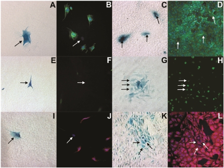
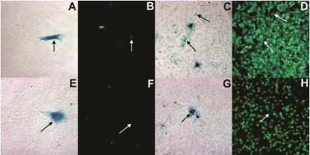
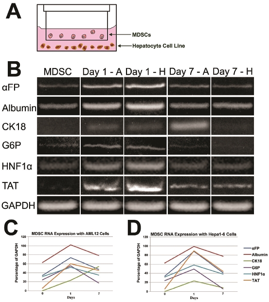
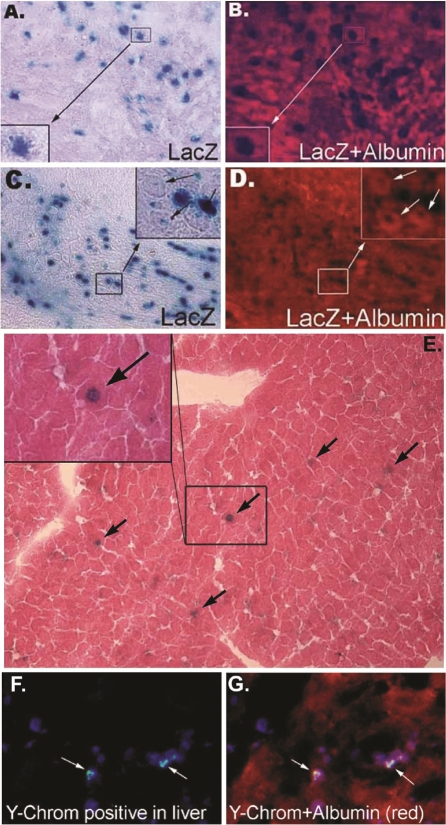
The liver is unique for its ability to regenerate after injury, however, critical injuries or disease cause it to lose this quality. Stem cells have been explored as a possibility to restore the function of seriously damaged livers, based on their self-renewability and multiple differentiation capacity. These experiments examine the ability of muscle derived stem cells (MDSCs) to differentiate into hepatocyte-like cells in vitro and acquire functional liver attributes for repairing damaged livers. In vitro experiments were performed using MDSCs from postnatal mice and mouse hepatocyte cell lines. Our data revealed that MDSCs differentiated into hepatocyte-like cells and expressed liver cell markers, albumin, hepatocyte nuclear factor 4 α, and alpha feto-protein, both at the RNA and protein level. Additionally, in vivo studies showed successful engraftment of MDSCs into hepatectomized mouse livers of mice. These results provide evidence suggesting that MDSCs have the capacity to differentiate into liver cell-like cells and may serve as potential candidates to aid in liver regeneration.

Keywords: Differentiation; Hepatectomy; Liver; Muscle Derived Stem Cells.


Figures

Figure 1
Figure 1
Bright field (A, C, E, G, I, K) and fluorescent microscopy (B, D, F, H, J, L) images demonstrating MDSCs differentiation into hepatocyte-like cells when co-cultured with the AML12 hepatocyte cell line after 1 day (A, B, E, F, I, J) and 7 days (C, D, G, H, K, L). In the bright field images, MDSCs are denoted by β-galactosidase expression (blue) and by black arrows whereas in fluorescent images, they are denoted by white arrows at the same location. In the fluorescent images, the co-cultured cells are stained for albumin (green: B, D), HNF4α (green: F, H) and αFP (red: J, L). The cell nuclei were stained with DAPI (blue).
Figure 2
Figure 2
Bright field (A, C, E, G) and fluorescent microscopy (B, D, F, H) images demonstrating MDSCs differentiation into hepatocyte-like cells when co-cultured with the Hepa1-6 hepatocyte cell line after 1 day (A, B, E, F) and 7 days (C, D, G, H). MDSCs are denoted by β-galactosidase expression (blue) and black arrows in bright field microscopy images. Fluorescent images, were taken at the exact location, where MDSCs are denoted by white arrows and stained for albumin (green: B, D) and HNF4α (red: F, H). The cell nuclei were stained with DAPI (blue).
Figure 3
Figure 3
Representation of the transwell co-culture setup where MDSCs were placed in the transwell and the hepatocyte cell lines (AML12 and Hepa1-6) were placed in the bottom of the multiwell dish. Gene expression profile of mouse MDSCs co-culture with either AML12 (Day 1 - A and Day 7 - A) or Hepa1-6 (Day 1 - H and Day 7 - H) hepatocyte cell lines using a transwell plate after 1 and 7 days (B). The expression of hepatocyte cell markers in MDSCs when co-cultured with AML12 (C) and Hepa1-6 (D) cell lines for different periods of time are shown with normalization to the loading control, GAPDH.
Figure 4
Figure 4
MDSCs differentiate into liver-like cells in vivo. Images of liver tissue harvested 3 days after MDSCs transplantation and stained for β-galactosidase (A, blue) or albumin (B, red). Similarly, liver tissue harvested 7 days after MDSC transplantation was also stained for β-galactosidase (C, blue) or albumin (D, red). Long term MDSCs engraftment into hepatectomized livers was observed after 3 months (E). As indicated by the black arrows, β-galactosidase positive (blue) MDSCs are found in the liver tissue subjected to hemotoxylin and eosin staining. A magnified image is shown in the inset of this figure. FISH analysis revealing the donor signal (Ychromosome) within injured liver tissue of female recipients, 7 days after male MDSCs were transplanted (F, G). MDSCs were stained for cell nuclei, blue; Y-chromosome, green; and albumin, red.

Similar articles

Cited by

References

    1. Ebrahimkhani MR, Elsharkawy AM, Mann DA. Wound healing and local neuroendocrine regulation in the injured liver. Expert Rev Mol Med. 2008;10:e11. - PubMed
    1. Alwayn IP, Verbesey JE, Kim S, Roy R, Arsenault DA, Greene AK, Novak K, Laforme A, Lee S, Moses MA, Puder M. A critical role for matrix metalloproteinases in liver regeneration. J Surg Res. 2008;145:192–198. - PMC - PubMed
    1. Michalopoulos GK, DeFrances MC. Liver regeneration. Science. 1997;276:60–66. - PubMed
    1. Iimuro Y, Nishio T, Morimoto T, Nitta T, Stefanovic B, Choi SK, Brenner DA, Yamaoka Y. Delivery of matrix metalloproteinase-1 attenuates established liver fibrosis in the rat. Gastroenterology. 2003;124:445–458. - PubMed
    1. Schuppan D, Afdhal NH. Liver cirrhosis. Lancet. 2008;371:838–851. - PMC - PubMed

Publication types

LinkOut - more resources