

Variations on theme: spindle assembly in diverse cells
- PMID: 20830494
- PMCID: PMC5290749
- DOI: 10.1007/s00709-010-0205-x
Variations on theme: spindle assembly in diverse cells
Abstract
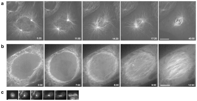
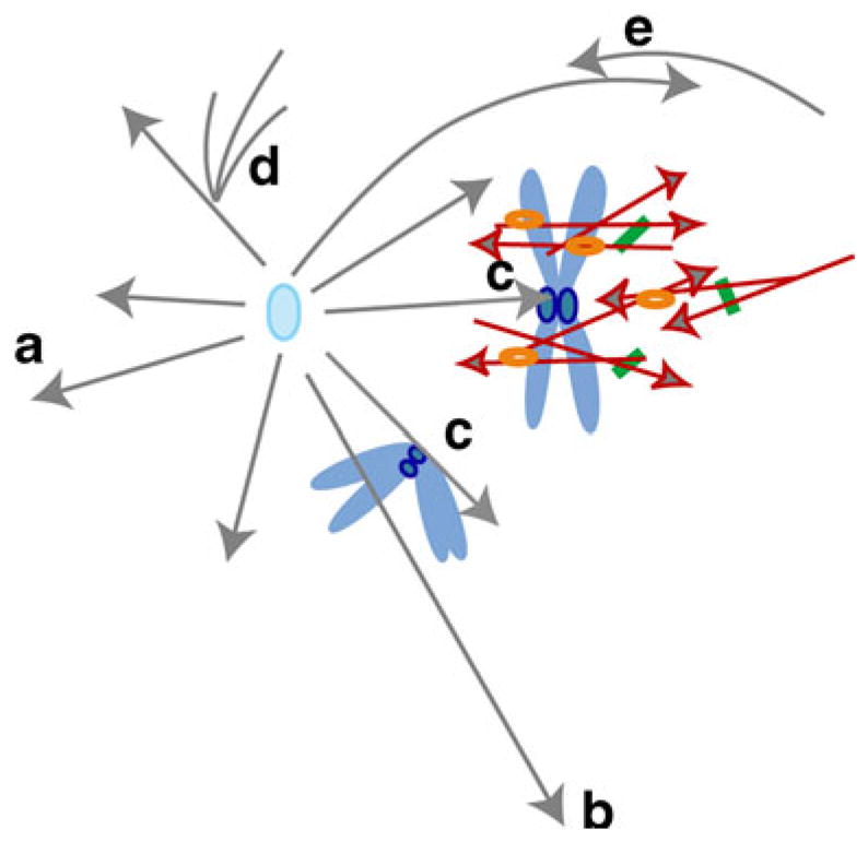
The mitotic spindle faithfully separates the genetic material, and has been reverently observed for well over a century. Across eukaryotes, while the mechanisms for moving chromosomes seem quite conserved, mechanisms for assembling the spindle often seem distinct. Two major pathways for spindle assembly are known, one based on centrosomes and the other based on chromatin, and these pathways are usually considered to be fundamentally different. We review observations of spindle assembly in animals, fungi, and plants, and argue that microtubule assembly at a particular location, centrosomes, or chromatin, reflects contingent, cell-type specific factors, rather than reflecting a fundamental distinction in the process of spindle building. We hypothesize that the essential process for spindle assembly is the motor-driven organization of microtubules that accumulate in the form of dense bundles at or near the chromosomes.
Conflict of interest statement
The authors declare that they have no conflict of interest.
Figures
Comment in
-
The same, but different--a bird's-eye view on mitosis.Protoplasma. 2011 Jul;248(3):437-8. doi: 10.1007/s00709-011-0299-9. Epub 2011 Jun 22. Protoplasma. 2011. PMID: 21695411 No abstract available.
References
-
- Ambrose JC, Cyr R. Mitotic spindle assembly and function. In: Verma DPS, Hong Z, editors. Cell division control in plants. Springer; Berlin: 2007. pp. 141–167.
-
- Ambrose JC, Cyr R. Mitotic spindle organization by the preprophase band. Mol Plant. 2008;1:950–960. - PubMed
-
- Bannigan A, Lizotte-Waniewski M, Riley M, Baskin TI. Emerging molecular mechanisms that power and regulate the anastral mitotic spindle of flowering plants. Cell Motil Cytoskel. 2008;65:1–11. - PubMed
-
- Baskin TI, Cande WZ. The structure and function of the mitotic spindle in flowering plants. Annu Rev Plant Physiol Plant Mol Biol. 1990;41:277–315.
Publication types
MeSH terms
Grants and funding
LinkOut - more resources
Full Text Sources
