. 2010 Oct 19;49(41):8892-901.
doi: 10.1021/bi1007723.

Use of recombinant cell-permeable small peptides to modulate glucocorticoid sensitivity of acute lymphoblastic leukemia cells

Affiliations

Use of recombinant cell-permeable small peptides to modulate glucocorticoid sensitivity of acute lymphoblastic leukemia cells

Chuan-dong Geng et al. Biochemistry. .

Abstract

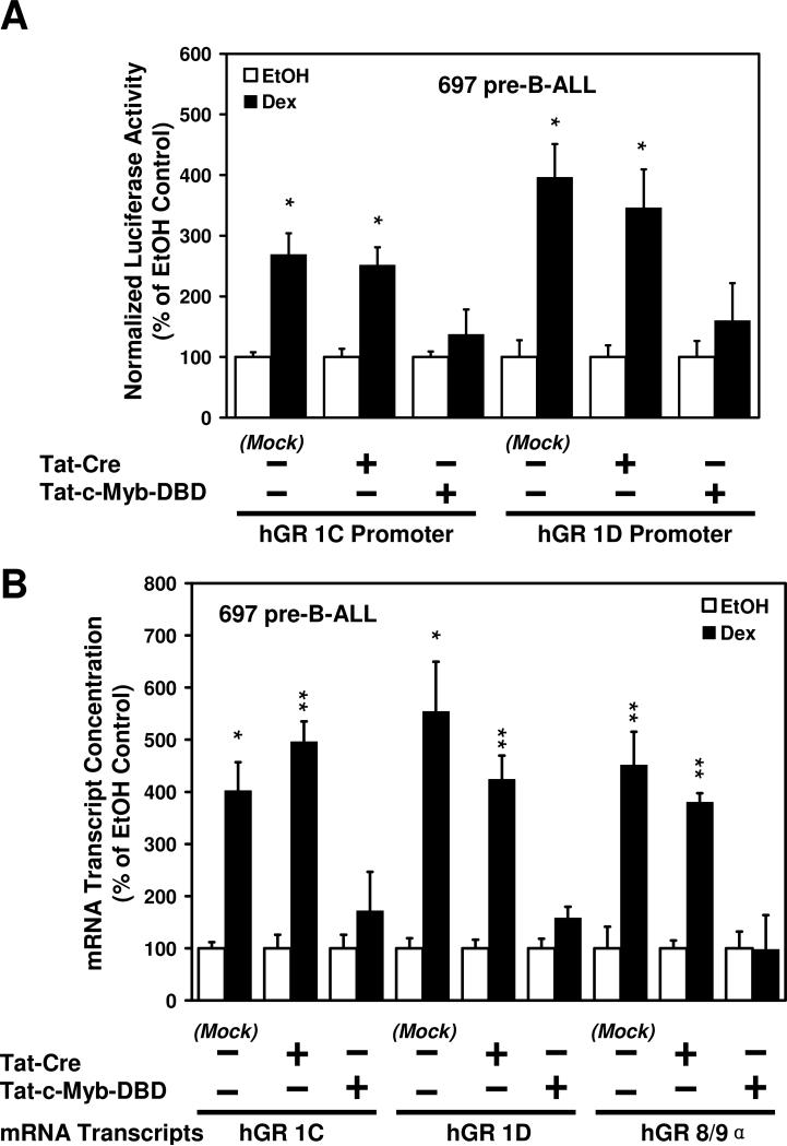
Glucocorticoid (GC) hormones induce apoptosis in T-cell and pre-B-cell acute lymphoblastic leukemia (ALL) cells. Steroid-mediated apoptosis requires a threshold level of the glucocorticoid receptor (GR) protein, and increasing the intracellular GR levels in ALL cells would augment their hormone sensitivity. A protein transduction domain (PTD) approach was used to accomplish this. We produced an HIV Tat PTD domain fusion protein (Tat-GR(554-777)) that potentially competes for the degradation of GR protein by the ubiquitin-proteasome system and should thus increase its intracellular levels by "stabilizing" the GR. We also designed a fusion peptide for the c-Myb DNA binding domain, Tat-c-Myb DBD, since the biological function of this peptide as a dominant negative inhibitor of the c-Myb protein was already known. Purified, bacterially expressed Tat-c-Myb DBD and Tat-GR(554-777) exhibited highly efficient transduction into cultured ALL cell lines including 697 (pre-B-ALL) and CEM-C7 (T-ALL) cells. As expected, the transduced Tat-c-Myb DBD peptide inhibited steroid-mediated stimulation of a GR promoter-luciferase reporter gene. Significantly, transduced Tat-GR(554-777) effectively increased intracellular GR levels in the GC-resistant T-ALL cell line, CEM-C1, and in the pre-B-ALL 697 cell line. Furthermore, transduction of Tat-GR(554-777) rendered GC-resistant CEM-C1 cells sensitive to steroid killing and further sensitized 697 cells to steroid. The use of Tat-fusion peptide transduction may eventually lead to innovative therapeutic modalities to improve the clinical response of patients suffering from T-cell and pre-B-cell acute lymphoblastic leukemia by increasing steroid responsiveness and perhaps converting steroid-resistant leukemia to a hormone-responsive phenotype.


Figures

FIGURE 1
FIGURE 1
Recombinant expression and purification of Tat-fusion proteins and peptides. The E. coli strain BL21 (DE3) Gold® carrying pTAT-vectors for fusion proteins or peptides was induced by IPTG to express the Tat-fusion protein as described in Methods and Materials. The baterial lysates were loaded on to a Ni-NTA column to purify 6 x His-tagged fusion proteins. The Ni-column purified proteins were loaded on to an ion-exchange column and eluted with increasing NaCl concentrations. A) diagram of the process to obtain purified recombinant Tat-fusion proteins or peptides; B) SDS-PAGE of protein aliquots from each step of purification of recombinant Tat-c-Myb-DBD. The SDS-gels were visualized by silver staining; C) Purified Tat-GR554-777 and Tat-Cre protein.
FIGURE 2
FIGURE 2
Transduction of Tat-fusion peptides into lymphoid cells. A) The purified recombinant Tat-fusion peptides ,Tat-c-Myb-DBD, Tat-GR554-777, Tat-Cre, and Tat-GR263-428, were labeled with FITC and incubated with cultured cells for 36 hours (697 pre-B-ALL cells) or 48 hours (CEM-C7 T-ALL cells). The samples were then observed via fluorescence microscopy. B) Reversibility of transduction of recombinant Tat-fusion peptides in lymphoid cells. The cultured cells were transduced with the Tat-GR554-777 fusion peptide for 48 hours, such that virtually 100% of the cells in the two cell lines contained the fluorescent peptide. The cells were washed and incubated in peptide-free medium for the indicated times. The FITC signal for the labeled peptide was gradually lost from the cells, indicating the reversibility of the process.
FIGURE 3
FIGURE 3
Transduction of the Tat-c-Myb-DBD peptide effectively suppresses auto-upregulation of GR gene expression in 697 pre-B-ALL cells. A) transient reporter gene analysis of hGR gene promoters in 697 cells transduced with the purified recombinant Tat-c-Myb-DBD peptide and the GR promoter luciferase reporter genes. For each sample, the GR-luciferase reporter gene plasmid was cotransfected with a CMV promoter-β-galactosidase reporter gene. The luminescent signal of the sample was normalized to the value obtained for the β-galactosidase activity in the same sample, to adjust for variations in transfection efficiencies between samples. The normalized values for the Dex-treated sample was divided by the normalized value from its respective ethanol control sample, and this value was multiplied by 100 to give the percent of ethanol control value.; B) QRT-PCR analysis of the expression of endogenous hGR trancripts in Tat-c-Myb DBD transduced 697 pre-B-ALL cells. Purified recombinant Tat-Cre was used as the control Tat-fusion peptide to treat 697 cells. Three separate experiments were performed. The data represent the mean ± the SEM for Dex-treated samples versus the EtOH treated control (value set at 100%). *: P<0.05 and **: P<0.01.
FIGURE 4
FIGURE 4
Transduction of the Tat-c-Myb-DBD peptide leads to the suppressed expression of intracellular GR protein levels in 697 pre-B-ALL cells. 697 cells were incubated with the Tat-c-Myb – DBD for 48 hours, after which they were treated for an additional 18 hours with 1 μM Dex or an equivalent volume of the ethanol vehicle. Western blotting was performed as described in Materials and Methods. GR levels were decreased in 697 cells as a result of Tat-c-Myb DBD transduction. GAPDH was used to monitor loading of total protein on the gel.
FIGURE 5
FIGURE 5
Transduction of Tat-GR554-777 effectively increases the intracellular GR protein levels in CEM-C1 T-ALL cells and 697 pre-B-ALL cells in a dose-dependent manner. After 48 hours of transduction, the expression of endogenous GR protein at each dose of transduced Tat-GR554-777 was detected by western blotting with a GR specific antibody that recognizes a sequence in the N-terminus (which is absent in Tat-GR554-777). The amount of GR protein was determined by densitometry and normalized to GAPDH protein levels in the same sample. The plot displays the fold differences comparing to the Tat-Cre-treated control cell samples (which are set at 1).
FIGURE 6
FIGURE 6
Sensitization of cells to glucocorticoid-induced cell death by transduction of purified recombinant Tat-GR554-777 into ALL cells. Cells were transduced with the indicated concentrations of Tat-GR554-777 for 48 hours. They were then treated for an additional 36 hours with 1 μM Dex or an equivalent volume of ethanol vehicle. Cell viability, determined using the Vybrant Apoptosis Assay Kit #4 (Invitrogen), in the Dex treated sample was compared to the ethanol vehicle-treated respective control sample. A) Transduction of Tat-GR554-777 results in conversion of the steroid-resistant CEM-C1 T-ALL cell to hormone-sensitivity in a dose dependent manner; B) Tat-GR554-777 further sensitizes the 697 pre-B-ALL to GC killing.
FIGURE 7
FIGURE 7
Transduction of Tat-GR554-777 specifically increases the intracellular GR protein levels in CEM-C1 T-ALL cells and 697 pre-B-ALL cells and sensitizes cells to glucocorticoid-induced cell death. A) and D)- The expression of the endogenous GR protein was detected by western blotting in ALL cells transduced by Tat-Cre, Tat-GR554-777 and Tat-GR263-428. A) CEM-C1 T-ALL cells; D) 697 pre-B-ALL cells. B) and E) The amount of GR protein in ALL cells transduced by recombinant Tat-fusion peptides was determined by densitometry and normalized to GAPDH protein levels. Figures show the fold differences of transduced samples versus the mock controls. B) CEM-C1 cells; E) 697 cells. C) and F) Cell cultures were transduced for 48 hours with the respective Tat-fusion peptide and then were treated for an additional 36 hours with 1 μM Dex or an equivalent volume of the ethanol vehicle. Cell viability, determined using the Vybrant Apoptosis Assay Kit #4 (Invitrogen), in the Dex treated sample was expressed as a percent of the respective ethanol vehicle-treated control sample. C) Tat-GR554-777 specifically results in conversion of the steroid-resistant CEM-C1 T-ALL cell to hormone-sensitivity; F) Tat-GR554-777, but not Tat-Cre or Tat-GR263-428, further sensitizes the 697 pre-B-ALL cells to GC killing.

Similar articles

Cited by

References

    1. Pulte D, Gondos A, Brenner H. Trends in 5- and 10-year survival after diagnosis with childhood hematologic malignancies in the United States, 1990-2004. J. Natl. Cancer Inst. 2008;100:1301–1309. - PubMed
    1. DeAngelo DJ. The treatment of adolescents and young adults with acute lymphoblastic leukemia. Hematology Am. Soc. Hematol. Educ. Program. 2005;2005:123–130. - PubMed
    1. Pui CH, Evans WE. Treatment of acute lymphoblastic leukemia. N. Engl. J. Med. 2006;354:166–178. - PubMed
    1. Pui CH, Relling MV, Downing JR. Acute lymphoblastic leukemia. N. Engl. J. Med. 2004;350:1535–1548. - PubMed
    1. Rivera GK, Pinkel D, Simone JV, Hancock ML, Crist WM. Treatment of acute lymphoblastic leukemia -- 30 years’ experience at St. Jude Children's Research Hospital. N. Engl. J. Med. 1993;329:1289–1295. - PubMed

Publication types

MeSH terms

Substances