Review
. 2010 Oct;162(1):12-23.
doi: 10.1111/j.1365-2249.2010.04210.x.

CD23/FcεRII: molecular multi-tasking

Affiliations
Review

CD23/FcεRII: molecular multi-tasking

M Acharya et al. Clin Exp Immunol. 2010 Oct.

Abstract

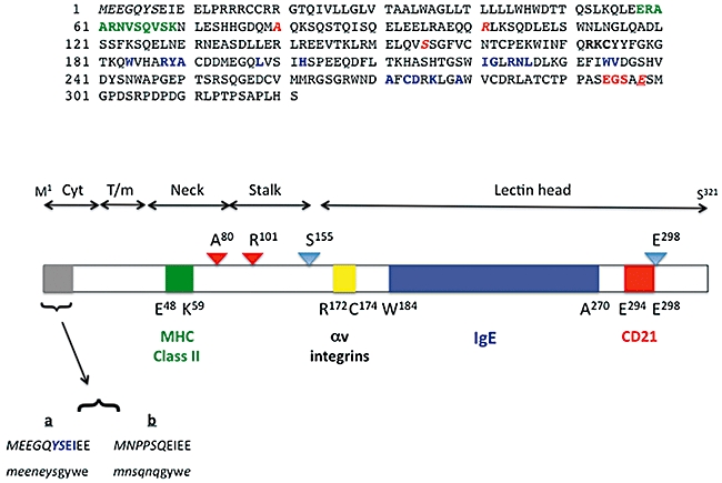
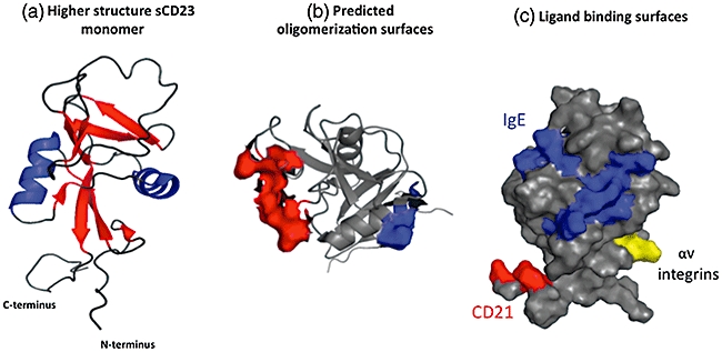
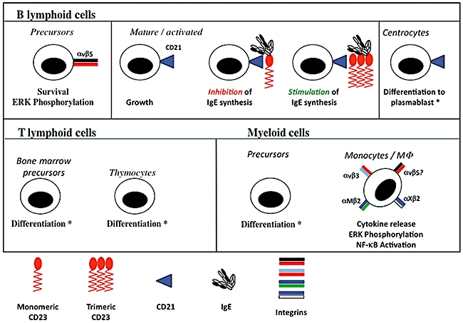
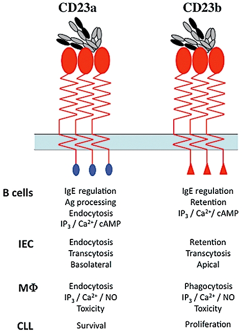
CD23 is the low-affinity receptor for immunoglobulin (Ig)E and plays important roles in the regulation of IgE responses. CD23 can be cleaved from cell surfaces to yield a range of soluble CD23 (sCD23) proteins that have pleiotropic cytokine-like activities. The regions of CD23 responsible for interaction with many of its known ligands, including IgE, CD21, major histocompatibility complex (MHC) class II and integrins, have been identified and help to explain the structure-function relationships within the CD23 protein. Translational studies of CD23 underline its credibility as a target for therapeutic intervention strategies and illustrate its involvement in mediating therapeutic effects of antibodies directed at other targets.


Figures

Fig. 1
Fig. 1
Primary structural features of human CD23. (a) The 321 amino acids that comprise the primary structure of human CD23a [1,2,55]. Individual contact residues or binding regions for CD23 ligands are shown in green (major histocompatibility complex class II [45]), bold type (αv integrins [51]), blue (IgE [44]) or red (CD21 [44]). The colours of the latter three interaction surfaces are identical to those used on the models shown in Fig. 3C. The unique N-terminal sequence of CD23a is shown as italicized letters, and protease target residues are shown as italicized red letters. Note that glu298 is underlined, as it is both a CD21 contact residue and a protease target [27,28,44]. (b) The rough delineation of regions of human CD23a into the lectin head, stalk, neck, transmembrane helix and cytoplasmic tail domains. The bar diagram shows the position of the ligand binding surfaces using the same colours used in (a) (and in Fig. 3c), with the exception of the αv integrin binding site, which is depicted in yellow; residues at the boundaries of the ligand interaction surfaces are noted. Protease binding sites are identified by triangles, with red indicating an ADAM cleavage site [24,25] and blue a der p1 target [27,28]; the proteases cleave to the C-terminal side of the indicated residues. Finally, the unique N-terminal sequences of the CD23a and CD23b isoforms for human (CAPITAL) and murine (lower-case) proteins are shown. The unique sequences are shown in italics and the common sequences contributed by exon 3 of the relevant gene are shown in normal case [55]. The motif responsible for endocytosis of human CD23a is highlighted in blue [79,80].
Fig. 3
Fig. 3
Higher structural features of CD23. (a) A ribbon diagram of the CD23 protein (left-hand image) illustrating the organization of the eight β strands and 2 α helices that comprise the lectin head domain; loop regions are also clear [43]. (b) Surface view of the CD23 lectin head demonstrating the position of acidic (red) and basic (blue) residues predicted to be important in oligomerization of CD23 [43]. (c) The spatial separation of the interaction surfaces for IgE (blue), CD21 (red) and αv integrins (yellow) on the lectin head domain [38,43]; these colours match those used in the primary structural diagram in Fig. 1. Note that the major histocompatibility complex class interaction site resides at the base of the stalk region and is not included in nuclear magnetic resonance (NMR) or crystal structures to date. Images were generated with Pymol software (DeLano Scientific LLC, San Francisco, CA, USA) using the NMR structure of CD23, accession code IT8C.
Fig. 2
Fig. 2
Pleiotropy of human sCD23. The effects of sCD23 on B cells, T cells and myeloid cells are illustrated showing biological responses and, where appropriate, signalling responses. An asterisk (*) indicates that the observed effect required the presence of interleukin (IL)-1α. Where defined, the receptors mediating the effects of sCD23 are identified (e.g. blue triangle for CD21, pairs of coloured bars for integrins). The upper panel shows the effects on B cell precursors [34,51,52], on activated B cells alone [30] and in the presence of immunoglobulin (Ig)E bound to monomeric and trimeric sCD23 [29], and on centrocytes [33]. The lower left-hand panel illustrates effects on CD4+ bone marrow and thymocyte [36] T cell precursors and the lower right-hand panel shows the responses driven by sCD23 in monocyte precursors [35] and mature monocyte/MΦ[38,47,49,50,61,62]. Note that no data confirming that αvβ5 can promote cytokine release are currently available, although this is highly likely, and the question-mark (?) on the figure denotes this.
Fig. 4
Fig. 4
Functions of CD23 isoforms. The functions of the two human CD23 isoforms are noted in the table below the sketches of CD23a (blue N-terminal domain) and CD23b (red) N-terminal domain. The isoforms are shown as trimeric forms interacting with a single IgE drawn to represent the highly bent conformation of IgE [127]. Isoform functions are described for B lymphocytes [55,60,64], intestinal epithelial cells (IEC) [14,79,80], monocyte/macrophages (MΦ) [49,50,61,64,123] and CLL cells [7].

Similar articles

Cited by

References

    1. Ludin C, Hofstetter H, Sarfati M, et al. Cloning and expression of the cDNA coding for a human lymphocyte IgE receptor. EMBO J. 1987;6:109–14. - PMC - PubMed
    1. Suter U, Bastos R, Hofstetter H. Molecular structure of the gene and the 5′-flanking region of the human lymphocyte immunoglobulin E receptor. Nucleic Acids Res. 1987;15:7295–308. - PMC - PubMed
    1. Conrad DH, Ford JW, Sturgill JL, Gibb DR. CD23: an overlooked regulator of allergic disease. Curr Allergy Asthma Rep. 2007;7:331–7. - PubMed
    1. Bonnefoy JY, Lecoanet-Henchoz S, Gauchat JF, et al. Structure and functions of CD23. Int Rev Immunol. 1997;16:113–28. - PubMed
    1. Gould HJ, Sutton BJ, Beavil AJ, et al. The biology of IGE and the basis of allergic disease. Annu Rev Immunol. 2003;21:579–628. - PubMed

Publication types