Comparative Study
. 2010 Sep 10:11:202.
doi: 10.1186/1471-2474-11-202.

Assessment of nerve involvement in the lumbar spine: agreement between magnetic resonance imaging, physical examination and pain drawing findings

Affiliations
Comparative Study

Assessment of nerve involvement in the lumbar spine: agreement between magnetic resonance imaging, physical examination and pain drawing findings

Bo C Bertilson et al. BMC Musculoskelet Disord. .

Abstract

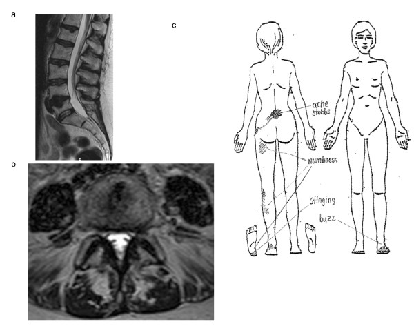
Background: Detection of nerve involvement originating in the spine is a primary concern in the assessment of spine symptoms. Magnetic resonance imaging (MRI) has become the diagnostic method of choice for this detection. However, the agreement between MRI and other diagnostic methods for detecting nerve involvement has not been fully evaluated. The aim of this diagnostic study was to evaluate the agreement between nerve involvement visible in MRI and findings of nerve involvement detected in a structured physical examination and a simplified pain drawing.

Methods: Sixty-one consecutive patients referred for MRI of the lumbar spine were - without knowledge of MRI findings - assessed for nerve involvement with a simplified pain drawing and a structured physical examination. Agreement between findings was calculated as overall agreement, the p value for McNemar's exact test, specificity, sensitivity, and positive and negative predictive values.

Results: MRI-visible nerve involvement was significantly less common than, and showed weak agreement with, physical examination and pain drawing findings of nerve involvement in corresponding body segments. In spine segment L4-5, where most findings of nerve involvement were detected, the mean sensitivity of MRI-visible nerve involvement to a positive neurological test in the physical examination ranged from 16-37%. The mean specificity of MRI-visible nerve involvement in the same segment ranged from 61-77%. Positive and negative predictive values of MRI-visible nerve involvement in segment L4-5 ranged from 22-78% and 28-56% respectively.

Conclusion: In patients with long-standing nerve root symptoms referred for lumbar MRI, MRI-visible nerve involvement significantly underestimates the presence of nerve involvement detected by a physical examination and a pain drawing. A structured physical examination and a simplified pain drawing may reveal that many patients with "MRI-invisible" lumbar symptoms need treatment aimed at nerve involvement. Factors other than present MRI-visible nerve involvement may be responsible for findings of nerve involvement in the physical examination and the pain drawing.


Figures

Figure 1
Figure 1
No MRI-visible nerve involvement though obvious physical examination findings. Figure 1ab: MRI of the lumbar spine of a woman aged 61 years. T2-weighted sagittal (a) and axial L4-5 (b) scans. The radiological assessment noted no visible nerve involvement but a slight paramedial disc protrusion at level L4-5, where an intraosseous disc hernia was also seen. Figure 1c: Pain drawing made by the patient in figure 1ab. The initial impression assessment of the pain drawing was that she had a left-sided L5 radiculopathy. Physical examination findings included: sensibility to touch and pain impaired in the lateral part of the left calf and slightly impaired in the whole left leg and lower left side of the trunk; tibialis posterior reflexes absent bilaterally and Achilles and patellar reflexes impaired bilaterally; motor function impaired for big toe extension and flexion on the left side. Patient history included 23 years of chronic backache, heel and Achilles pain (left side) and also urinary incontinence. Symptoms were initially acute when she fell from 3 meters and landed on her back. Standing, lifting, sitting and other axial loading of the spine increased her symptoms. Lying down relieved her symptoms.
Figure 2
Figure 2
MRI visible nerve involvement though no physical examination findings. Figure 2abc: MRI of the lumbar spine of a man aged 48 years. T2-weighted sagittal right foraminal (a), sagittal right lateral (b) and axial L5-S1 (c) scans. The radiological assessment noted visible nerve involvement bilaterally at L5 and right-sided nerve involvement at S1 due to disc hernia and intervertebral arthrosis. Figure 2d: Pain drawing made by the patient in figure 2abc. The initial impression assessment of the pain drawing was that he had right-sided L4-S1 radiculopathy. No pathological findings were observed in the physical examination except that the right plantar reflex was slightly impaired. Patient history included 9 years of walking difficulties but no back pain. Symptoms started when he carried a heavy weight. Walking or standing still for 5 minutes made his right leg cramp and feel numb, like "lots of lactic acid". Bending forward relieved the pain.

Similar articles

Cited by

References

    1. Dworkin RH, O'Connor AB, Backonja M, Farrar JT, Finnerup NB, Jensen TS, Kalso EA, Loeser JD, Miaskowski C, Nurmikko TJ. et al.Pharmacologic management of neuropathic pain: evidence-based recommendations. Pain. 2007;132(3):237–251. doi: 10.1016/j.pain.2007.08.033. - DOI - PubMed
    1. Haswell K, Gilmour J, Moore B. Clinical decision rules for identification of low back pain patients with neurologic involvement in primary care. Spine. 2008;33(1):68–73. doi: 10.1097/BRS.0b013e31815e3949. - DOI - PubMed
    1. Haefeli M, Elfering A. Pain assessment. Eur Spine J. 2006;15(Suppl 1):S17–24. doi: 10.1007/s00586-005-1044-x. - DOI - PMC - PubMed
    1. Koes BW, van Tulder MW, Ostelo R, Kim Burton A, Waddell G. Clinical guidelines for the management of low back pain in primary care: an international comparison. Spine. 2001;26(22):2504–2513. doi: 10.1097/00007632-200111150-00022. discussion 2513-2504. - DOI - PubMed
    1. Melton L. Taking a shot at neuropathic pain. The Lancet Neurology. 2003;2 - PubMed

Publication types

LinkOut - more resources