

The proliferation, apoptosis, invasion of endothelial-like epithelial ovarian cancer cells induced by hypoxia
- PMID: 20831794
- PMCID: PMC2944817
- DOI: 10.1186/1756-9966-29-124
The proliferation, apoptosis, invasion of endothelial-like epithelial ovarian cancer cells induced by hypoxia
Abstract
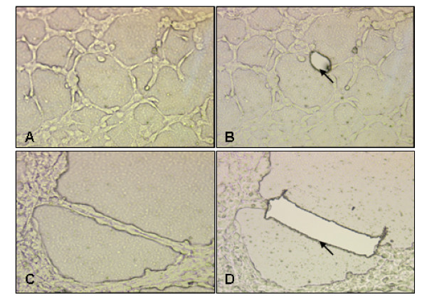
Background: Epithelial ovarian cancer is one of the most malignant cancers in women because metastasis occurs in the most of patients by the time of diagnosis. Cancer cells have strong capacity to form angiogenesis or vasculogenic mimicry, which plays the major role in its malignant phenotype. Vasculogenic mimicry might contribute to the failure of the angiogenesis-targeted therapy strategies. Under the microenvironment of the tumor, hypoxia is the most common phenomena because of the vast energy and oxygen consuming. In the present study, the endothelial-like cells induced by hypoxia from SKOV-3 and ES-2 ovarian cancer cells were harvested to investigate the changes in their biological behaviors.
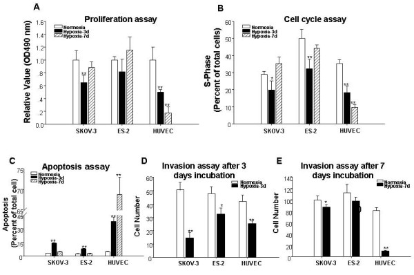
Methods: The endothelial-like cells from SKOV-3 and ES-2 cells were harvested by laser capture microdissection. The biological behaviors of the endothelial-like cells, including proliferation, cell cycle, apoptosis, invasion and telomerase activity were determined by MTT, FCM, Transwell chamber and TRAP-ELISA methods. HIF-1α is the most important factor for the behavior changes under hypoxic condition. Some other genes relative to biological behaviors are also changes following the changes of HIF-1α. In order to elucidate the underlying mechanisms for these changes by hypoxia, the relative genes expressions including HIF-1α, CyclinD1, Flk-1, VEGF, p53 and V-src were determined by real-time PCR.
Results: SKOV-3 and ES-2 cells were resistant to hypoxia by adoption of proliferation, apoptosis, differentiation and invasion. Combined with other studies, the more poorly cancer cells differentiate, the more strongly cells are resistant to hypoxia, the more possible to form vasculogenic mimicry. The changes in the expression of HIF-1α, and HIF-1α-dependent VEGF, Flk-1, Cyclin D1, and HIF-1α-independent p53 have been involved in this process.
Conclusions: HIF-1α took an important role in the behavioral changes of SKOV-3 and ES-2 cells by hypoxia. At the same time, other mechanisms were also involved in this process.
Figures

References
-
- Huang S, Robinson JB, Deguzman A, Bucana CD, Fidler IJ. Blockade of nuclear factor-kappaB signaling inhibits angiogenesis and tumorigenicity of human ovarian cancer cells by suppressing expression of vascular endothelial growth factor and interleukin 8. Cancer Res. 2000;60:5334–5339. - PubMed
-
- Demeter A, Varkonyi T, Csapo Z, Szantho A, Olah J, Papp Z. [Assessment of prognostic factors in common ovarian tumors of varying malignancy] Magy Onkol. 2004;48:259–265. - PubMed
Publication types
MeSH terms
Substances
LinkOut - more resources
Full Text Sources
Medical
Research Materials
Miscellaneous
