. 2010 Sep 10:3:21.
doi: 10.1186/1757-2215-3-21.

Leucine-rich alpha-2-glycoprotein-1 is upregulated in sera and tumors of ovarian cancer patients

Affiliations

Leucine-rich alpha-2-glycoprotein-1 is upregulated in sera and tumors of ovarian cancer patients

John D Andersen et al. J Ovarian Res. .

Abstract

Background: New biomarkers that replace or are used in conjunction with the current ovarian cancer diagnostic antigen, CA125, are needed for detection of ovarian cancer in the presurgical setting, as well as for detection of disease recurrence. We previously demonstrated the upregulation of leucine-rich alpha-2-glycoprotein-1 (LRG1) in the sera of ovarian cancer patients compared to healthy women using quantitative mass spectrometry.

Methods: LRG1 was quantified by ELISA in serum from two relatively large cohorts of women with ovarian cancer and benign gynecological disease. The expression of LRG1 in ovarian cancer tissues and cell lines was examined by gene microarray, reverse-transcriptase polymerase chain reaction (RT-PCR), Western blot, immunocytochemistry and mass spectrometry.

Results: Mean serum LRG1 was higher in 58 ovarian cancer patients than in 56 healthy women (89.33 ± 77.90 vs. 42.99 ± 9.88 ug/ml; p = 0.0008) and was highest among stage III/IV patients. In a separate set of 193 pre-surgical samples, LRG1 was higher in patients with serous or clear cell ovarian cancer (145.82 ± 65.99 ug/ml) compared to patients with benign gynecological diseases (82.53 ± 76.67 ug/ml, p < 0.0001). CA125 and LRG1 levels were moderately correlated (r = 0.47, p < 0.0001). LRG1 mRNA levels were higher in ovarian cancer tissues and cell lines compared to their normal counterparts when analyzed by gene microarray and RT-PCR. LRG1 protein was detected in ovarian cancer tissue samples and cell lines by immunocytochemistry and Western blotting. Multiple iosforms of LRG1 were observed by Western blot and were shown to represent different glycosylation states by digestion with glycosidase. LRG1 protein was also detected in the conditioned media of ovarian cancer cell culture by ELISA, Western blotting, and mass spectrometry.

Conclusions: Serum LRG1 was significantly elevated in women with ovarian cancer compared to healthy women and women with benign gynecological disease, and was only moderately correlated with CA125. Ovarian cancer cells secrete LRG1 and may contribute directly to the elevated levels of LRG1 observed in the serum of ovarian cancer patients. Future studies will determine whether LRG1 may serve as a biomarker for presurgical diagnosis, disease recurrence, and/or as a target for therapy.


Figures
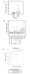
Figure 1
Figure 1
ELISA detection of serum LRG1. A) Serum LRG1 concentrations were determined for 58 ovarian cancer patients and 56 of the control patients. Box plots are presented here; the solid line indicates the median serum LRG1 for each group. Serum levels of LRG1 were significantly higher in the ovarian cancer sera than in control sera, after adjusting for age (p=0.0008). B) LRG1 in serum of individual patients with benign and malignant gynecological diseases. Median LRG1 values for each group are indicated by the solid bars. Dashed line indicates the mean LRG1 concentration from control serum in panel A. LRG1 concentrations are significantly higher in serum of women with ovarian cancer (serous and clear cell subtypes) than in serum of women with other gynecological diseases (p <0.0001). C) Receiver operator curves (ROC) for CA125 alone (blue line), LRG1 alone (red line) and LRG1 in combination with CA125 (green line). The area under the curve (AUC) for CA125 alone was 0.88, for LRG1 alone the AUC = 0.77, and the AUC for CA125 and LRG1 together was 0.89. There was no significant difference in sensitivity between CA125 alone and CA125 in combination with LRG1 (p=0.2728).
Figure 2
Figure 2
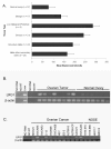
Expression of LRG1 transcripts in ovarian cancer tissues and cell lines. A) Microarray analysis of LRG1 gene expression in ovarian cancer tissues was performed on Affymetrix HU_133 gene chips. Mean expression of LRG1 RNA was determined for normal ovary, benign ovary tumors, and primary and metastatic ovarian cancers. (n) = number of samples per tissue type. B) RT-PCR of LRG1 expression in ovarian cancer tissue samples (N = 8) relative to normal ovary tissue (N =7). C) LRG1 expression in ovarian cancer cell lines compared to immortalized NOSE cell lines. β-actin was used as an amplification control.
Figure 3
Figure 3
Expression and localization of LRG1 protein in ovarian cancer tissues and cell lines. A) 50 µg of total protein extract from ovarian cancer tissues (N = 7) and normal ovaries (N =5) were evaluated by Western blot for LRG1 protein expression. Kidney was used as a positive control tissue, as it contains an abundance of epithelial cells. B) LRG1 protein expression in 20 µg of total protein extract from ovarian cancer cell lines and immortalized NOSE cells. β-actin was used as the loading control. C) Left panel; silver stained polyacrylamide gel of LRG1 purified from human plasma, PNGase F, and purified LRG1 treated with PNGase F. Right panel; Western blot for LRG1 in protein extracts from cell lines with and without PNGase F treatment. D) Subcellular localization of LRG1 is shown by immunocytochemistry in ovarian cancer cell lines (OVCAR5, OVCAR433, OV2008, C-13, and SKOV3) and immortalized NOSE cell line (1816-575); 200X magnification, scale bar = 20 µm. FITC (green) = LRG1, DAPI (blue) = nucleus.
Figure 4
Figure 4
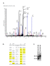
Secretion of LRG1 into conditioned media by ovarian cancer cell line NIH:OVCAR5. A) MS spectrum for LRG1 peptide, ALGHLDSGNR, identified in the spent media of NIH:OVCAR5 cells with 95% (peptide) probability. Conditioned media from the ovarian cancer cell line was concentrated and processed for MSMS analysis. LRG1was identified with low (protein) probability with a single peptide in the complex mixture. The identity of the peptide was confirmed by manual inspection. Peak assignments are indicated. B) m/z for predicted b- and y- ions for peptide ALGHLDSGNR. Highlighted peaks were identified in the spectrum shown in A. C) Western immunoblot of conditioned media from NOSE cell line 1816-575 and ovarian cancer cell line NIH:OVCAR5. 50 µg of concentrated, conditioned media from each cell line was loaded. Position of molecular weight standards, left.

References

    1. Ries L, Harkins D, Krapcho M, Mariotto A, Miller BA, Feuer EJ, Clegg L, Eisner MP, Horner MJ, Howlader N, Hayat M, Hankey BF, Edwards BK. SEER Cancer Statistics Review, 1975-2003. Book SEER Cancer Statistics Review, 1975-2003 (Editor ed.^eds.). City. 2005.
    1. Jemal A, Siegel R, Ward E, Hao Y, Xu J, Murray T, Thun MJ. Cancer statistics, 2008. CA Cancer J Clin. 2008;58:71–96. doi: 10.3322/CA.2007.0010. - DOI - PubMed
    1. Badgwell D, Bast RC Jr. Early detection of ovarian cancer. Dis Markers. 2007;23:397–410. - PMC - PubMed
    1. Yurkovetsky ZR, Linkov FY, D EM, Lokshin AE. Multiple biomarker panels for early detection of ovarian cancer. Future Oncol. 2006;2:733–741. doi: 10.2217/14796694.2.6.733. - DOI - PubMed
    1. Fung ET. A recipe for proteomics diagnostic test development: the OVA1 test, from biomarker discovery to FDA clearance. Clin Chem. pp. 327–329. - DOI - PubMed