. 2010 Sep 11:7:25.
doi: 10.1186/1743-8977-7-25.

Role of surface charge and oxidative stress in cytotoxicity of organic monolayer-coated silicon nanoparticles towards macrophage NR8383 cells

Affiliations

Role of surface charge and oxidative stress in cytotoxicity of organic monolayer-coated silicon nanoparticles towards macrophage NR8383 cells

Sourav Bhattacharjee et al. Part Fibre Toxicol. .

Abstract

Background: Surface charge and oxidative stress are often hypothesized to be important factors in cytotoxicity of nanoparticles. However, the role of these factors is not well understood. Hence, the aim of this study was to systematically investigate the role of surface charge, oxidative stress and possible involvement of mitochondria in the production of intracellular reactive oxygen species (ROS) upon exposure of rat macrophage NR8383 cells to silicon nanoparticles. For this aim highly monodisperse (size 1.6 ± 0.2 nm) and well-characterized Si core nanoparticles (Si NP) were used with a surface charge that depends on the specific covalently bound organic monolayers: positively charged Si NP-NH2, neutral Si NP-N3 and negatively charged Si NP-COOH.

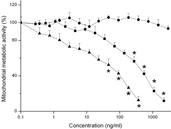
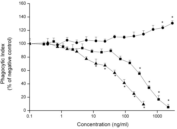
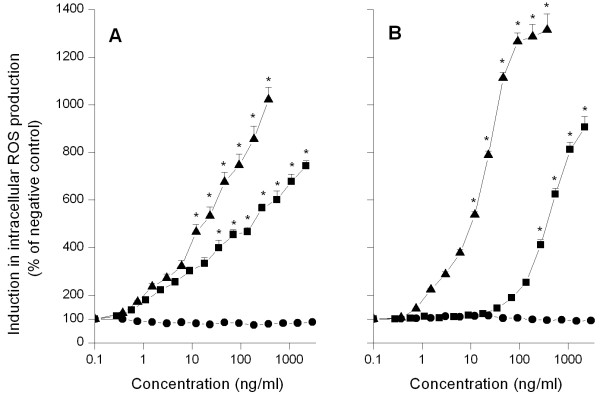
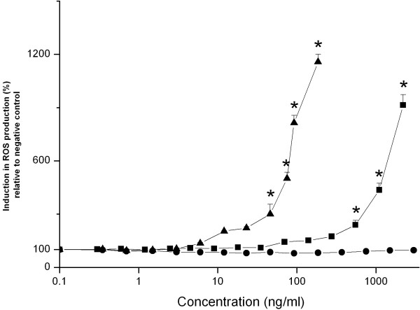
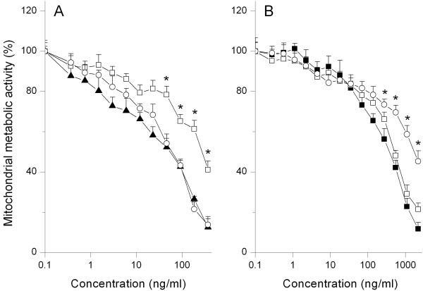
Results: Positively charged Si NP-NH2 proved to be more cytotoxic in terms of reducing mitochondrial metabolic activity and effects on phagocytosis than neutral Si NP-N3, while negatively charged Si NP-COOH showed very little or no cytotoxicity. Si NP-NH2 produced the highest level of intracellular ROS, followed by Si NP-N3 and Si NP-COOH; the latter did not induce any intracellular ROS production. A similar trend in ROS production was observed in incubations with an isolated mitochondrial fraction from rat liver tissue in the presence of Si NP. Finally, vitamin E and vitamin C induced protection against the cytotoxicity of the Si NP-NH2 and Si NP-N3, corroborating the role of oxidative stress in the mechanism underlying the cytotoxicity of these Si NP.

Conclusion: Surface charge of Si-core nanoparticles plays an important role in determining their cytotoxicity. Production of intracellular ROS, with probable involvement of mitochondria, is an important mechanism for this cytotoxicity.


Figures

Figure 1
Figure 1
Effect of 24 hours exposure of NR8383 cells to increasing concentrations of Si NP-NH2 (black triangle), Si NP-N3 (black square) and Si NP-COOH (black dot) on mitochondrial metabolic activity (measured by the MTT assay). Error bars show standard error of mean (n = 3). The asterisk (*) sign signifies P < 0.05.
Figure 2
Figure 2
Effect of 24 hours exposure of NR8383 cells to increasing concentrations of Si NP-NH2 (black triangle), Si NP-N3 (black square) and Si NP-COOH (black dot) on phagocytosis (measured as the phagocytic index). Error bars show standard error of mean (n = 3). The asterisk (*) sign signifies P < 0.05.
Figure 3
Figure 3
Induction of intracellular ROS production (measured using the DCFH-DA assay) in NR8383 (A) and Caco-2 (B) cells after 24 hours exposure to increasing concentrations of Si NP-NH2 (black triangle), Si NP-N3 (black square) and Si NP-COOH (black dot). Error bars show standard error of mean (n = 3). The asterisk (*) sign signifies P < 0.05.
Figure 4
Figure 4
Induction of ROS production (measured using the DCFH-DA assay) upon 90 minutes incubations of an isolated rat liver mitochondrial fraction with increasing concentrations of Si NP-NH2 (black triangle), Si NP-N3 (black square) and Si NP-COOH (black dot). Error bars show standard error of mean (n = 3). The asterisk (*) sign signifies P < 0.05.
Figure 5
Figure 5
Protective effect of preincubation of NR8383 cells with vitamin E (open square) and vitamin C (open circle) against cytotoxicity (measured using the MTT assay) detected after 24 hours exposure to serial dilutions of (A) Si NP-NH2 (black triangle) and (B) Si NP-N3 (black square). Error bars show standard error of mean (n = 3). The asterisk (*) sign signifies P < 0.05 (compared to data without antioxidants).

References

    1. Derfus AM, Chan WCW, Bhatia SN. Probing the cytotoxicity of semiconductor quantum dots. Nano Lett. 2004;4:11–18. doi: 10.1021/nl0347334. - DOI - PMC - PubMed
    1. Klauser F, Stijepovic R, Endstrasser N, Jaksch S, Memmel N, Scheier P. Oxidation study of silicon nanoparticle thin films on HOPG. Surf Sci. 2009;603:2999–3004. doi: 10.1016/j.susc.2009.08.007. - DOI
    1. Park J, Gu L, Maltzahn GV, Ruoslahti E, Bhatia SN, Sailor MJ. Biodegradable luminescent porous silicon nanoparticles for in vivo applications. Nat Mater. 2009;8(4):331–6. doi: 10.1038/nmat2398. - DOI - PMC - PubMed
    1. Burns AA, Vider J, Ow H, Herz E, Penate-Medina O, Baumgart M, Larson SM, Wiesner U, Bradbury M. Fluorescent silica nanoparticles with efficient urinary excretion for nanomedicine. Nano Lett. 2009;9(1):442–448. doi: 10.1021/nl803405h. - DOI - PMC - PubMed
    1. Rosso-Vasic M, Sprujit E, van Lagen B, De Cola L, Zuilhof H. Alkyl-functionalized oxide-free silicon nanoparticles: synthesis and optical properties. Small. 2008;4(10):1835–1841. doi: 10.1002/smll.200800066. - DOI - PubMed

Publication types

LinkOut - more resources