Review
. 2010 Oct;22(5):640-50.
doi: 10.1016/j.ceb.2010.08.019. Epub 2010 Sep 9.

Cellular mechanisms regulating epithelial morphogenesis and cancer invasion

Affiliations
Review

Cellular mechanisms regulating epithelial morphogenesis and cancer invasion

Ryan S Gray et al. Curr Opin Cell Biol. 2010 Oct.

Abstract

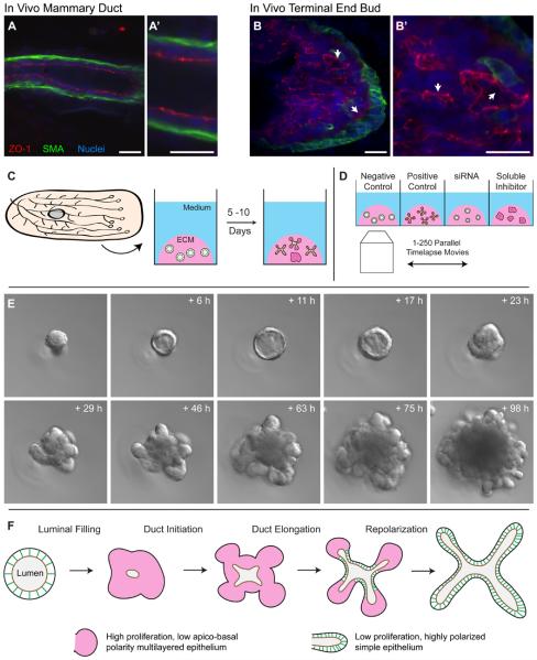
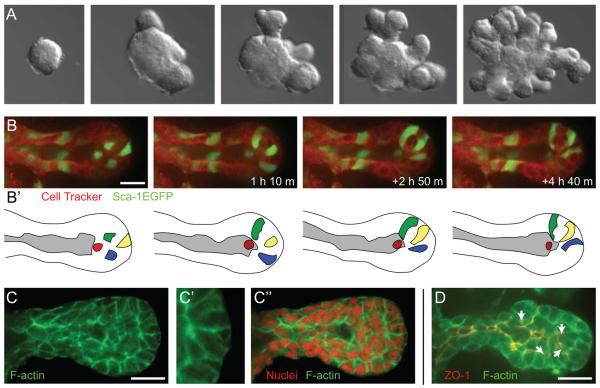
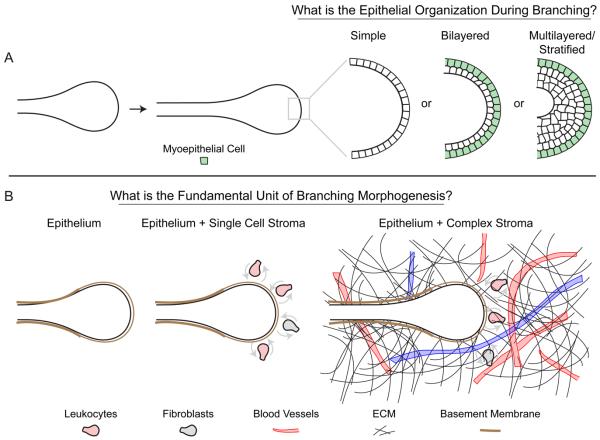
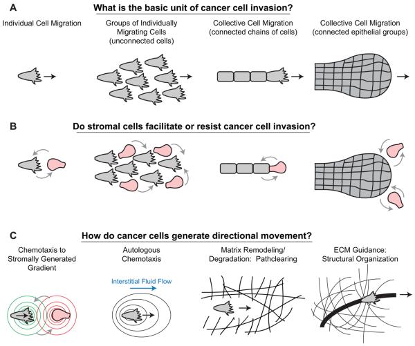
The cellular mechanisms driving mammalian epithelial morphogenesis are of significant fundamental and practical interest. Historically, these processes have been difficult to study directly, owing to the opacity and relative inaccessibility of mammalian tissues. Recent experimental advances in timelapse imaging and in 3D organotypic culture have enabled direct observation of epithelial morphogenesis. In the mammary gland, branching morphogenesis is observed to proceed through a novel form of collective epithelial migration. The active unit of morphogenesis is a multilayered epithelium with reduced apico-basal polarity, within which cells rearranged vigorously. From within this multilayered state, new ducts initiate and elongate into the matrix without leading cellular extensions or dedicated leaders. We discuss the implications of these findings on our understanding of epithelial morphogenesis in other organs and in cancer progression.


Figures

Figure 1
Figure 1. 3D primary organotypic culture makes mammary branching morphogenesis observable
(A, B) Confocal images of anti-body stained sectioned in vivo mammary duct (A) and terminal endbud (B), illustrating the normal in vivo composition of apical tight junctions (ZO-1; red) and the basal myoepithelial cell layer (SMA; green). (C) The mammary “organoid” assay involves isolation of epithelial fragments each initially consisting of 100-500 cells (“organoids”) from mammary glands through a combination of mechanical and enzymatic disruption [14,15,35]. These organoids are then subsequently embedded in Matrigel – a laminin, collagen IV, heparin sulfate rich matrix- and fed a serum-free media containing defined growth factors (e.g. FGF2). Fragments then develop over a period of 5-10 days and undergo a complex program of branching morphogenesis. (D) Mice can yield 1-5,000 organoids each, thereby enabling organoids from the same mouse to be cultured in different microenvironmental conditions [13,14]. The large scale enables parallel experimental design in which epithelium from the same mouse is allocated to different ECM, solution or perturbation (e.g. siRNA) conditions and enables the consequences of these variables to be assessed individually or parametrically with genetically identical starting material. Advances in timelapse imaging automation make it possible to image 100-500 movies in parallel and monitor individual cell behaviors within branching mammalian epithelia over 10s to 100s of hours. (E) Organoids first clear their lumen to a simple, bilayered architecture, then fill their lumen with cells prior to initiating, elongating and bifurcating new ducts. Images courtesy of Kim-Vy Nguyen-Ngoc, Johns Hopkins Medical School. (F) Mammary branching morphogenesis involves large, but transient, changes in apico-basal polarity, proliferation and epithelial organization. The functional unit of morphogenesis has high proliferation, low polarity and multilayered organization (pink). As elongation ceases the epithelium reorganizes and restores highly polarized simple epithelial organization (green). Images from A-B are reprinted with permission from [10]. Scale bars are 20 microns.
Figure 2
Figure 2. The basic program of mammary branching morphogenesis
(A) Epithelial fragments (“organoids”) isolated from the mammary gland will undergo a complex program of branching morphogenesis in 3D ECM culture. This program involves the de novo initiation, elongation and bifurcation of new ducts. (B-B′) Cells within the elongation front rearrange vigorously and there do not appear to be dedicated leader cells [14]. (C-C″) Elongating mammary ducts have a multilayered organization at the elongation front and do not have actin based subcellular protrusions into the ECM [14]. (D) Within the multilayered endbud there is typically a tight junction (ZO-1) lined main lumen as well as isolated microlumens within the multilayered region (arrows) [14]. ECM = extracellular matrix, ZO-1 = zona occludens 1, a tight junction marker. Images from B-D are reprinted with permission from [10]. Scale bars are 20 microns.
Figure 3
Figure 3. Conserved developmental mechanisms for branching morphogenesis
(A) Most mammalian epithelial tubes have a simple organization. However, during active morphogenesis both the mammary [14] and salivary [26] epithelia are multilayered or stratified. By contrast lung appears to retain a simple organization during branching morphogenesis [40]. (B) During branching morphogenesis in each of the epithelial organs there is a rich diversity of ECM proteins and stromal cell types, including diverse leukocytes and fibroblasts. Functional evidence has been provided for the importance of macrophages during mammary development [22,26], but it remains a relatively open question which stromal cell types are required for, or assist in, the normal development of the epithelial organs. ECM = extracellular matrix.
Figure 4
Figure 4. Cellular mechanisms driving epithelial cancer invasion
(A) Cancer cell invasion has been observed to proceed by both individual and collective mechanisms. Collective migration strategies can include: chains, files and connected, largely epithelial groups such as pushing boundaries [52]. In addition, single cell invasion can involve large numbers of individually migrating cells, but the term collective cell migration is reserved for connected groups of cells. (B) Stromal cells, including macrophages [58] and fibroblasts [69], have been observed to assist in the invasion of tumors. It remains an open question which stromal cell types in vivo serve to assist or resist the invasion and dissemination of cancer cells. Stromal cells could influence the invasion of singly or collectively migrating cancer cells. (C) A diversity of mechanisms have also been reported to explain the guidance of cancer cell invasion, including: reciprocal gradients of EGF and CSF-1 between macrophages and cancer cells [58], autologous chemotaxis whereby molecules released by cancer cells are shifted towards the lymphatics by interstitial fluid flow [60], pathclearing through the ECM, for example by fibroblasts ahead of squamous cell carcinoma cells [69] or alternately reorganization of the ECM to promote the invasion of cancer cells [63]. ECM = extracellular matrix.

Similar articles

Cited by

References

    1. Sternlicht MD, Kouros-Mehr H, Lu P, Werb Z. Hormonal and local control of mammary branching morphogenesis. Differentiation. 2006;74:365–381. - PMC - PubMed
    1. Hennighausen L, Robinson GW. Information networks in the mammary gland. Nat Rev Mol Cell Biol. 2005;6:715–725. - PubMed
    1. Howlin J, McBryan J, Martin F. Pubertal mammary gland development: insights from mouse models. J Mammary Gland Biol Neoplasia. 2006;11:283–297. - PubMed
    1. Sternlicht MD. Key stages in mammary gland development: the cues that regulate ductal branching morphogenesis. Breast Cancer Res. 2006;8:201. - PMC - PubMed
    1. Hinck L, Silberstein GB. Key stages in mammary gland development: the mammary end bud as a motile organ. Breast Cancer Res. 2005;7:245–251. - PMC - PubMed

Publication types