. 2010 Sep 10;39(5):689-99.
doi: 10.1016/j.molcel.2010.08.008.

An Smc3 acetylation cycle is essential for establishment of sister chromatid cohesion

Affiliations

An Smc3 acetylation cycle is essential for establishment of sister chromatid cohesion

Frederic Beckouët et al. Mol Cell. .

Abstract

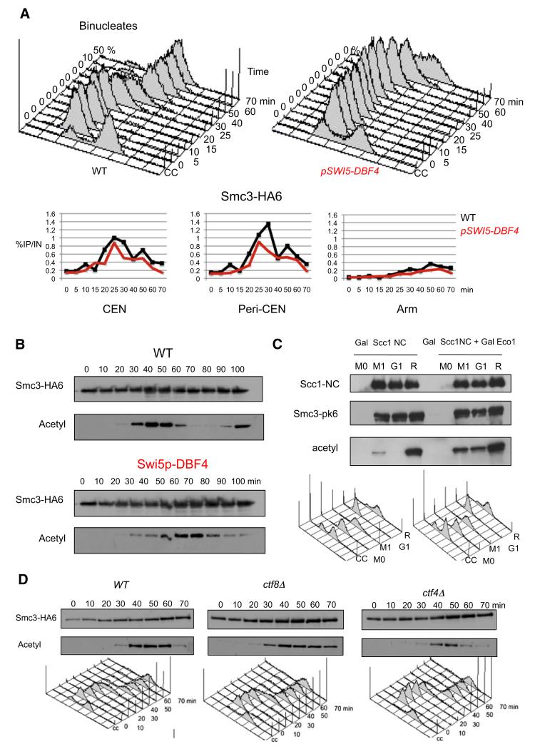
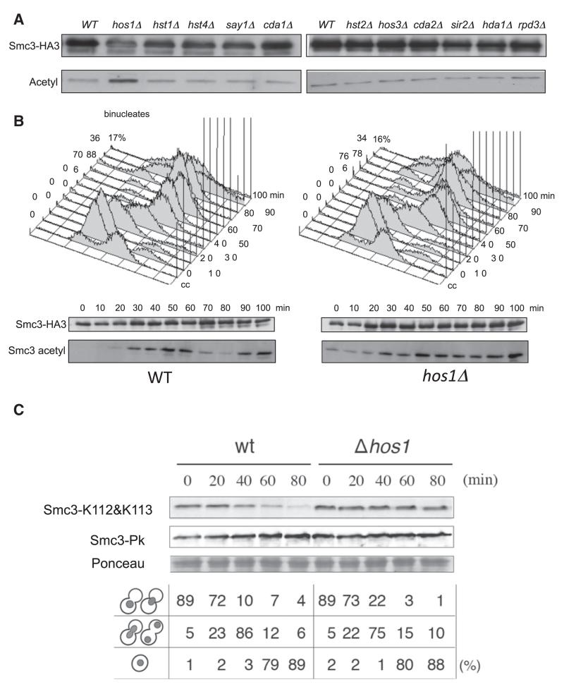
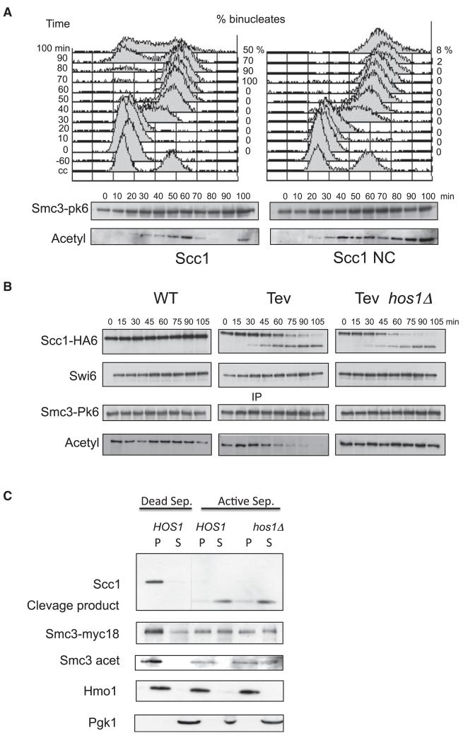
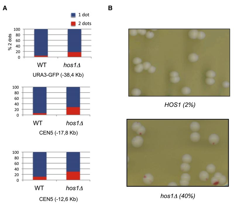
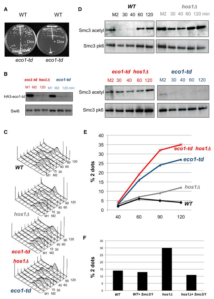
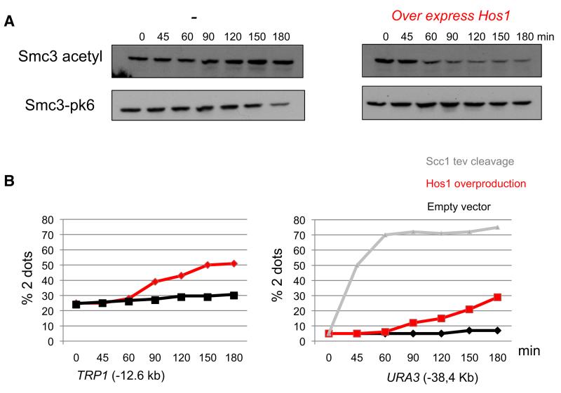
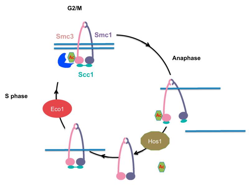
Sister chromatid cohesion is thought to involve entrapment of sister DNAs by a tripartite ring composed of the cohesin subunits Smc1, Smc3, and Scc1. Establishment of cohesion during S phase depends on acetylation of Smc3's nucleotide-binding domain (NBD) by the Eco1 acetyl transferase. It is destroyed at the onset of anaphase due to Scc1 cleavage by separase. In yeast, Smc3 acetylation is reversed at anaphase by the Hos1 deacetylase as a consequence of Scc1 cleavage. Smc3 molecules that remain acetylated after mitosis due to Hos1 inactivation cannot generate cohesion during the subsequent S phase, implying that cohesion establishment depends on de novo acetylation during DNA replication. By inducing Smc3 deacetylation in postreplicative cells due to Hos1 overexpression, we provide evidence that Smc3 acetylation contributes to the maintenance of sister chromatid cohesion. A cycle of Smc3 NBD acetylation is therefore an essential aspect of the chromosome cycle in eukaryotic cells.


Figures

Figure 1
Figure 1. Efficient Smc3 Acetylation by Eco1 at the Time of S Phase Depends on DNA Replication
(A) HA6-tagged Smc3 associated with three chromosomal loci (as measured by ChIP and qPCR) in K16655 (MATa SMC3-HA6) and K16653 (MATa SMC3-HA6, dbf4::SWI5p-DBF4) cells that are released from α factor-induced G1 arrest into fresh medium at 25°C. Cell cycle progression was monitored by flow cytometry of DNA content, and the time of anaphase was microscopically determined by the occurrence of binucleated cells (upper panel). An untagged strain (K699) yielded no significant ChIP-qPCR signal. (B) Smc3-HA6 was immunoprecipitated from wild-type (K16655) or SWI5p-DBF4 (K16653) cells that were released from α factor-induced G1 arrest into fresh medium at 25°C. The acetylation status of Smc3 was analyzed by western blotting using the α-acetyl lysine antibody (ST1027, Calbiochem). (C). Strains K17957 (MATa SMC3-PK6::KanMX4, TRP1::PMET3CDC20, pGAL1-10SCC1(R180D, R268D)-HA3::LEU2, his3::tetR-GFP::HIS3, YEplac195) K17955 (MATa SMC3-PK6::KanMX4, TRP1:: PMET3CDC20, pGAL1-10SCC1(R180D,R268D)-HA3:: LEU2, YEplac195-Gal-Eco1) were grown to log phase in synthetic medium lacking methionine and arrested in metaphase for 2 hr in YEP medium supplemented with 2 mM methionine and 2% raffinose. After 2 hr, the sample was divided into three subsamples, one being maintained in metaphase arrest, the second being released to G1 arrest, and the third being released to the next S phase. In all cases 2% galactose was added to the media for 80 min to induce expression of Scc1-NC-HA3. Samples were taken at the indicated times for preparation of whole cell extract. Scc1-HA3 was immunoprecipitated using α-HA antibody, and the acetylation level of co-immunoprecipitated Smc3 was subsequently measured by α-Smc3-acetyl-K113 (Figure S1) anti-body via western blotting. (D) Smc3-HA6 was immunoprecipitated from K16655 (MATa SMC3-HA6), K16842 (MATa SMC3-HA6 ctf8Δ), or K16845 (MATa SMC3-HA6 ctf4Δ) cells that were released from α factor-induced G1 arrest into fresh medium at 25°C. Western blots were performed using either HA-specific or acetyl-lysine-specific antibodies (ST1027, Calbiochem). Cell cycle progression was monitored by flow cytometry of DNA content, and the time of anaphase was microscopically determined by the occurrence of binucleated cells.
Figure 2
Figure 2. The Histone Deacetylase Hos1 Acts on Smc3
(A) Smc3-HA3 was immunopurified from 13 deacetylase deletion mutants, and the level of acetylated Smc3 was evaluated by western blotting using α-acetyl lysine antibody (ST1027, Calbiochem). (B) Smc3 acetylation pattern in K15544 (WT) and K16351 (hos1 Δ) cells that are released from α factor-induced G1 arrest (160 min) into fresh medium at 25°C. Smc3-HA3 was immunopurified from aliquots of the culture at the indicated time points, and Smc3 acetylation was analyzed by western blotting using a specific α-acetylated K113 antibody. Cell cycle progression was monitored by flow cytometry of DNA content, and the time of anaphase was microscopically determined by the occurrence of binucleated cells. (C) Strains ST231 (HOS1) and ST432 (hos1 Δ) growing in YPD medium were arrested at 23°C in G2/M phases for 2.5 hr after adding benomyl (80 μg/ml). Cells were then washed with YPD, re-suspended in YPD containing α factor, and cultured for 80 min at 23°C. The acetylation status and the level of Smc3-pk6 at the indicated time points were monitored by western blotting of crude extracts using α-pk- and α-Smc3-acetylated antibodies.
Figure 3
Figure 3. Hos1 Acts on Smc3 Released from Chromatin after Scc1 Cleavage
(A) Strain K16548 (MATa, SMC3-PK6, pGAL1-10-SCC1-HA3::LEU2) expresses Scc1-HA3 epitope from the GAL1-10 promoter. Strain K16546 (MATa, SMC3-PK6, pGAL1-10-SCC1(R180D, R268D)-HA3::LEU2) expresses a separase non-cleavable version of Scc1-HA3 from the GAL1-10 promoter. Both strains were arrested at 30°C in G1 by α factor in YEP medium containing 2% raffinose. After 1 hr in G1 phase, the GAL1-10 promoter was induced by addition of galactose. One hour after induction, cells were released from G1 arrest and samples were taken every 10 min for preparation of whole cell extract. Cell cycle progression was monitored by flow cytometry of DNA content and microscopic analysis of binucleate (anaphase) cells (upper panel). Cell extracts were prepared from aliquots of the culture at the indicated times and Smc3-PK6 immunoprecipitated using an antibody raised against the pk epitope tag. The acetylation level of Smc3 was monitored by western blotting using the α-acetyl lysine antibody (ST1027, Calbiochem). FACScan analysis shows that cells were arrested and released efficiently (upper panel). (B) Yeast strains K16394 (MATα PMET3CDC20, SCC1-HA6, SMC3-PK6, YEp-PGAL1TEV), K16397 (MATα PMET3CDC20, SCC1(TEV220)-HA6, SMC3-PK6, YEp-PGAL1TEV) and K16465 (MATα PMET3CDC20, SCC1(TEV220)-HA6 SMC3-PK6 hos1Δ, YEp-PGAL1TEV) were grown to log phase in synthetic medium lacking methionine and arrested in metaphase for 2 hr in YEP medium supplemented with 2 mM methionine and 2% raffinose (0 min). Expression of TEV protease was induced by addition of 2% galactose (0-105 min). Smc3-PK6 was immunoprecipitated and the level of Smc3 acetylation was monitored using lysine-specific antibodies (ST1027, Calbiochem). Cleavage of Scc1-HA6 was monitored by probing whole cell extracts against the HA epitopes. Swi6 serves as a loading control. (C) Chromatin isolated from the strain K17304 (MATa SCC1-HA6, SMC3-PK6, hos1Δ) arrested in nocodazole was incubated for 15 min at 25°C with a mixture of extracts from the mitotic strains K8965 (MATa esp1-1, pGAL1-10-ESP1), K17305 (MATa esp1-1, hos1 Δ, pGAL1-10-ESP1) and K8967 (MATa esp1-1, pGAL1-10-esp1-C1531A) overproducing wild-type (ESP1) or catalytically dead (esp1-C1531A) separase. Reactions were analyzed by western blotting using α-Myc, α-HA and α-Smc3 acetylated antibodies. Hmo1 and cytosolic pGK1 served as a loading control.
Figure 4
Figure 4. Cohesion Defect in hos1Δ
(A) Cells carrying MET-CDC20 and GFP labels at various loci grown to log phase in synthetic medium lacking methionine were transferred to YPD plus methionine. After 150 min, the percentages of separated GFP foci were scored. The tet operators are integrated at: URA3-GFP (38.4 kb to left of CEN5) in wild-type (K16472) and hos1 Δ (K16470) (upper panel); 17.8CEN6-GFP (17.8 kb to left of CEN5) in wild-type (K17508) and hos1 Δ (K17506) (middle panel); and −12.6CEN5-GFP (12.6 kb to left of CEN5) in wild-type (K17512) and hos1 Δ (K17510) (lower panel). (B) Sectoring assay for the chromosome loss using the SUP11-marked in HOS1 strain (K7040) and its hos1 Δ derivative (K16466).
Figure 5
Figure 5. Smc3 Needs to be Acetylated Specifically in S Phase to be Competent for Establishment of Cohesion
Exponential phase cells of strains K17515 (WT, PMET3CDC20, SMC3-PK6), K17516 (hos1 Δ, PMET3CDC20, SMC3-PK6), and K17376 (td-Eco1, PMET3CDC20, SMC3-PK6) and K17382 (td-Eco1 hos1 Δ, PMET3CDC20, SMC3-PK6) were grown to log phase in synthetic medium lacking methionine and arrested in metaphase for 2 hr in YEP medium supplemented with 2 mM methionine, 2% raffinose, and 2% galactose (M1). Eco1 was degraded by shifting cultures to degron-mediated proteolysis conditions (YEP raff/gal, 20 μg/ml doxycycline, at 37°C) for 90 min (M2). Subsequently, the cells were released from G2/M arrest to raff/gal media (lacking methionine) containing 20 μg/ml doxycycline. Thirty minutes after release at 37°C, nocodazole was added to arrest the cells again in G2/M phase. (A) Strains 16354 (WT) and 16353 (Eco1-deg) were grown at 25°C for 2 days on YP plates containing 2% raffinose and 2% galactose, then transferred to 37°C on plates containing 2% raffinose, 2% galactose, and doxycycline (5 μg/ml). (B) Western blots show degradation of endogenous HA-tagged td-Eco1 proteins (C) Fluorescence-activated cell sorting analysis shows that the release from Cdc20 arrest was complete and that all strains had completed DNA replication after 90 min. (D) Smc3-PK6 was immunopurified, and the level of acetylated Smc3 was evaluated by western blotting using α-acetyl lysine antibody (ST1027, Calbiochem). (E) Cohesion was measured by scoring the percentages of separated GFP dots at the TRP1 locus (12.6 kb from CEN4). (F) K17987 (hos1 Δ, 256LacO::TRP1, PMET3CDC20, YEp-SMC1/SMC3) K17988 (hos1 Δ, 256LacO::TRP1, PMET3CDC20, YEp), K17989 (256LacO::TRP1, PMET3CDC20, YEp-SMC1/SMC3), and K17990 (256LacO::TRP1, PMET3CDC20) were grown to log phase in synthetic medium lacking methionine and uracil and arrested in metaphase for 2 hr in YEP medium supplemented with 2 mM methionine. Cohesion defect was measured by scoring the percentages of separated GFP dots at the TRP1 locus (12.6 kb from CEN4).
Figure 6
Figure 6. Acetylation Stabilizes Cohesion
Strains K17513 (256LacO::TRP1, PMET3CDC20, YEp-PGAL1HOS1, SMC3-PK6), K17514 (256La-cO::TRP1, PMET3CDC20, YEp-PGAL1, SMC3-PK6), K17383 (tetO112::URA3, PMET3CDC20, YEp-PGAL1HOS1), K17384 (tetO112::URA3, MET3-CDC20, YEp-PGAL1), and K9027 (MATα, te-tO112::URA3, PMET3CDC20, scc1Δ, SCC1-TEV268-HA3, GAL-TEV) were grown to log phase in synthetic medium lacking methionine and arrested in metaphase for 2 hr in YEP medium sup-plemented with 2 mM methionine and 2% raffinose (0 min). Expression of Hos1 was induced by addition of 2% galactose (0-180 min). (A) Proteins from the strains K17513 and K17514 were extracted, and the levels of Smc3 acetylation and Smc3-PK6 at the indicated time points were monitored by western blotting of crude extracts using α-pk and α-acetylated Smc3 antibodies. (B) Cohesion was measured by scoring the percentages of separated GFP dots at the TRP1 (12.6 kb from CEN4) and URA3 (38.4 kb from CEN5) loci.
Figure 7
Figure 7. Smc3 Acetylation Cycle
During S phase, Eco1 acetylates Smc3. This reaction is required for the establishment of sister chromatid cohesion. In G2/M phases, cohesion has been established, and we propose that Smc3 acetylation may act to stabilize it. At the metaphase-to-anaphase transition, separase cleavage of Scc1 allows Hos1 to deacetylate Smc3.

Comment in

References

    1. Rolef Ben-Shahar T, Heeger S, Lehane C, East P, Flynn H, Skehel M, Uhlmann F. Eco1-dependent cohesin acetylation during establishment of sister chromatid cohesion. Science. 2008;321:563–566. - PubMed
    1. Ciosk R, Shirayama M, Shevchenko A, Tanaka T, Toth A, Shevchenko A, Nasmyth K. Cohesin’s binding to chromosomes depends on a separate complex consisting of Scc2 and Scc4 proteins. Mol. Cell. 2000;5:243–254. - PubMed
    1. Davie JK, Edmondson DG, Coco CB, Dent SY. Tup1-Ssn6 interacts with multiple class I histone deacetylases in vivo. J. Biol. Chem. 2003;278:50158–50162. - PubMed
    1. Gambus A, van Deursen F, Polychronopoulos D, Foltman M, Jones RC, Edmondson RD, Calzada A, Labib K. A key role for Ctf4 in coupling the MCM2-7 helicase to DNA polymerase alpha within the eukaryotic replisome. EMBO J. 2009;28:2992–3004. - PMC - PubMed
    1. Gruber S, Haering CH, Nasmyth K. Chromosomal cohesin forms a ring. Cell. 2003;112:765–777. - PubMed

Publication types

MeSH terms

LinkOut - more resources