. 2010 Sep 10;39(5):761-72.
doi: 10.1016/j.molcel.2010.08.013.

Loss of miR-200 inhibition of Suz12 leads to polycomb-mediated repression required for the formation and maintenance of cancer stem cells

Affiliations

Loss of miR-200 inhibition of Suz12 leads to polycomb-mediated repression required for the formation and maintenance of cancer stem cells

Dimitrios Iliopoulos et al. Mol Cell. .

Abstract

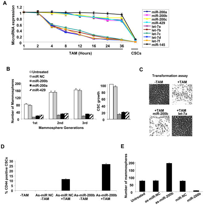
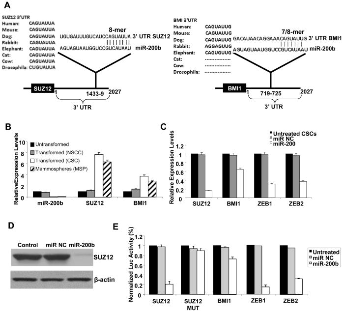
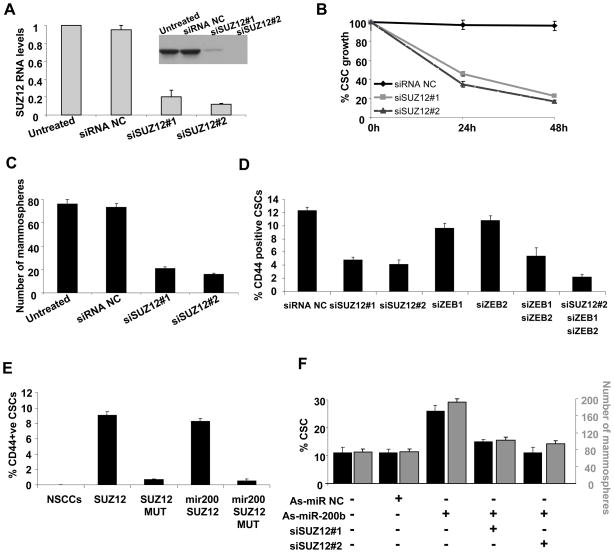
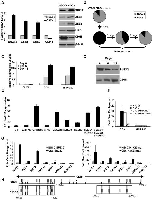
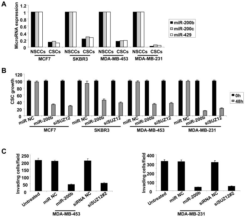
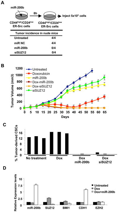
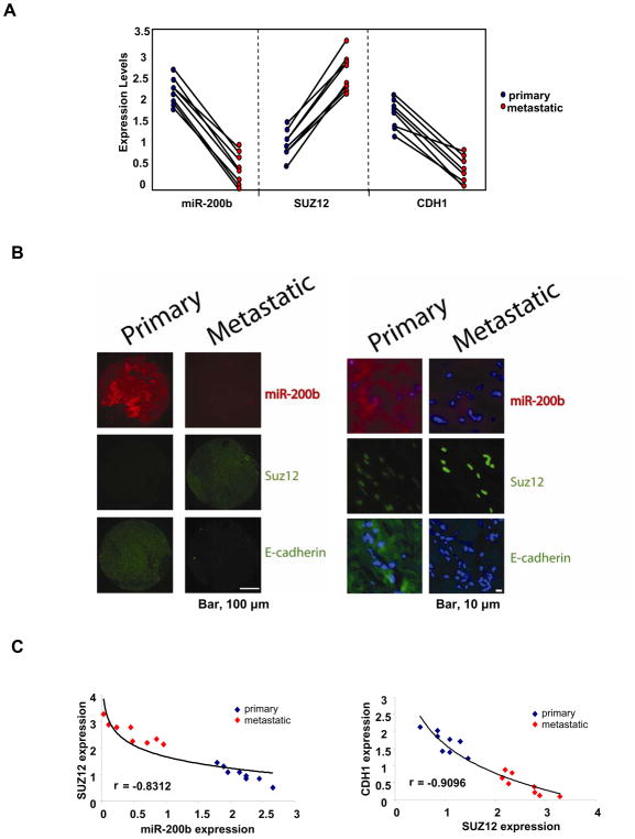
In an inducible oncogenesis model, the miR-200 family is inhibited during CSC formation but not transformation, and inhibition of miR-200b increases CSC formation. Interestingly, miR-200b directly targets Suz12, a subunit of a polycomb repressor complex (PRC2). Loss of miR-200 during CSC formation increases Suz12 expression, Suz12 binding, H3-K27 trimethylation, and Polycomb-mediated repression of the E-cadherin gene. miR-200b expression or Suz12 depletion blocks the formation and maintenance of mammospheres, and in combination with chemotherapy suppresses tumor growth and prolongs remission in mouse xenografts. Conversely, ectopic expression of Suz12 in transformed cells is sufficient to generate CSCs. The miR-200b-Suz12-cadherin pathway is important for CSC growth and invasive ability in genetically distinct breast cancer cells, and its transcriptional signature is observed in metastatic breast tumors. The interaction between miR-200 and Suz12 is highly conserved, suggesting that it represents an ancient regulatory mechanism to control the growth and function of stem cells.


Figures

Figure 1
Figure 1. MiR-200b Regulates Cancer Stem Cell Growth
(A) Expression (mean ± SD) of the indicated microRNAs at the indicated times after induction of transformation with tamoxifen (TAM) and in CSCs purified by flow cytometry. (B) CSCs and 1st, 2nd, 3rd passage mammospheres were transfected with the indicated microRNAs and examined for the percentage of CSC growth (relative to untransfected CSCs) and number of mammospheres/1000 cells. (C) Transformation of TAM-treated ER-Src cells transfected with miR-200b or let-7a microRNAs. Data are presented as mean ± SD. (D) Flow cytometry analysis of CD44/CD24 profiles and percentage of CSCs (mean ± SD) in the population (mean ± SD) after transfection with the indicated anti-sense microRNAs. (E) Number of mammospheres/1000 cells ((mean ± SD) after transfection of the indicated antisense microRNAs.
Figure 2
Figure 2. Direct Targeting of Suz12 by MiR-200b Through an Evolutionarily Conserved Interaction
(A) Predicted miR-200b binding sites in the 3′UTRs of Suz12 and Bmi1 with sequence complementarity and phylogenic conservation of 8-nt seed sequence indicated. Zeb1 and Zeb2 show similar complementarity as Suz12 with the exception of Drosophila. (B) MiR-200b, Suz12 and Bmi1 RNA levels (mean ± SD) in untransformed, transformed non-stem cancer cells (NSCCs), CSCs, and mammospheres (MSP). (C) RNA levels (mean ± SD) of the indicated genes after transfection with miR-200b or control microRNAs in CSCs. (D) Suz12 and β-actin protein levels after transfection with miR-200b or control microRNAs in CSCs. (E) Relative luciferase activity (mean ± SD) mediated by reporter constructs harboring the 3′UTR of the indicated genes (and mutated version of Suz12) upon transfection with miR-200b or control microRNAs.
Figure 3
Figure 3. Suz12 is Required for CSC and Mammosphere Growth Mediated by Loss of MiR-200
(A) Suz12 RNA and protein (inset) levels (mean ± SD) in CSCs treated with 2 different siRNAs against Suz12 or control siRNA (NC). (B) Percentage of CSCs obtained by sorting ER-Src cells treated with TAM for 36h were transfected with Suz12 or control siRNAs, and examined for the percentage of CSC growth (relative to untransfected cells) 24h and 48h afterwards. (C) Number of mammospheres/1000 cells (mean ± SD) 48h after transfection with Suz12 or control siRNAs. (D) Percentage CSCs (mean ± SD) of ER-Src transformed cells after transfection with siRNAs against the indicated genes. (E) NSCCs obtained by sorting an ER-Src transformed cell population were transfected with Suz12 or control expression vectors in the presence or absence of co-transfected miR-200 were analyzed for the percentage of CSCs by flow cytometry (mean ± SD). (F) Percentage of CSCs (black) and number of mammospheres (gray) after transfection with antisense-miR negative control (as-miR NC), antisense-miR-200b (as-miR-200b), or Suz12 siRNAs. Data are shows as mean ± SD.
Figure 4
Figure 4. MiR-200b Functions through Suz12 and H3K27 Tri-methylation in CSCs
(A) Suz12 and E-cadherin (Cdh1) mRNA (left) and protein (right) levels (mean ± SD) in NSCCs and CSCs. (B) Percentage (mean ± SD) of CSCs (black) and NSCCs (gray) at the indicated times after plating CSCs (obtained from sorting ER-Src transformed cells) under differentiation conditions. (C) Suz12, Cdh1, and miR-200 RNA levels (mean ± SD) during differentiation of CSCs. (D) Suz12 and Cdh1 protein levels during differentiation of CSCs. (E) Cdh1 RNA levels (mean ± SD) in CSCs after transfection of miR-200b or siRNAs against the indicated genes. (F) Chromatin immunoprecipitation showing association of Suz12 at CDH1 and control HNRPA2 gene in NSCCs and CSCs expressing miR-200 or a control microRNA. Data are presented as mean ± SD. (G) Suz12 association and H3-K27 trimethylation (mean ± SD) at the indicated loci in NSCCs and CSCs. (H) DNA methylation analysis of regions within the CDH1 promoter in CSCs and NSCCs. For each horizontal line, open circles indicate non-methylated residues and black circles indicated methylated residues in a given clone that was sequenced after bisulfite conversion.
Figure 5
Figure 5. MiR-200b-Suz12 Pathway Affects Growth and Invasive Ability of CSCs Derived from Genetically Distinct Breast Cancer Cell Lines
(A) MiR-200b, miR-200a and miR-429 expression levels in NSCCs and CSCs cells derived from the indicated breast cancer cell lines. (B) Growth of CSCs derived from the indicated cell lines 48h post transfection with miR-200b or siRNA against Suz12. Data are presented as mean ± SD. (C) Invasion assays in CSCs derived from MDA-MB-435 and MDA-MB-231 cells 12h post transfection with miR-200b or siRNA against Suz12. Data are presented as mean ± SD.
Figure 6
Figure 6. MiR-200b Expression or Suz12 Inhibition in Combination with Chemotherapy Prevents Tumor Relapse
(A) Tumor incidence in nude mice injected with CSCs from ER-Src transformed cells that were pre-treated with miR-200b or siRNA against Suz12 for 8h. (B) Tumor volume (mm3) in xenografts treated with doxorubicin, miR-200b, siSuz12#2 and combinations at days 10, 15, 20, and 25. (C) Percentage of CSCs derived from ER-Src tumors treated with doxorubicin combined with miR-200b or siSuz12. (D) Expression of the indicated RNAs (mean ± SD) in CD44high/CD24low cells taken from tumors at day 25 treated as indicated.
Figure 7
Figure 7. MiR-200b-Suz12-Cdh1 pathway in primary and metastatic human tumors
(A) MiR-200b, Suz12 and Cdh1 expression levels (mean ± SD) in primary and metastatic breast tumor pairs. (B) Microscopic images showing miR-200b RNA (in situ hybridization) as well as Suz12 and Cdh1 protein levels (immunohistochemistry) in primary and metastatic breast tumor pairs. Higher magnification shows the miR-200b cytoplsmic localization (red), Suz12 nuclear localization (green) and Cdh1 cytoplasmic localization (green); DAPI (blue) stained the nuclei. (C) Correlation between miR-200b and Suz12 expression and Cdh1 and Suz12 expression in primary and metastatic breast tumors.

Comment in

References

    1. Adam L, Zhong M, Choi W, Qi W, Nicoloso M, Arora A, Calin G, Wang H, Siefker-Radtke A, McConkey D, et al. miR-200 expression regulates epithelial-to-mesenchymal transition in bladder cancer cells and reverses resistance to epidermal growth factor receptor therapy. Clin Cancer Res. 2009;15:5060–5072. - PMC - PubMed
    1. Ailles LE, Weissman IL. Cancer stem cells in solid tumors. Curr Opin Biotechnol. 2007;18:460–466. - PubMed
    1. Aziz N, Cherwinski H, McMahon M. Complementation of defective colony-stimulating factor 1 receptor signaling and mitogenesis by Raf and v-Src. Mol Cell Biol. 1999;19:1101–1115. - PMC - PubMed
    1. Bao S, Tang F, Li X, Hayashi K, Gillich A, Lao K, Surani MA. Epigenetic reversion of post-implantation epiblast to pluripotent embryonic stem cells. Nature. 2009;461:1292–1295. - PMC - PubMed
    1. Ben-Porath I, Thomson MW, Carey VJ, Ge R, Bell GW, Regev A, Weinberg RA. An embryonic stem cell-like gene expression signature in poorly differentiated aggressive human tumors. Nat Genet. 2008;40:499–507. - PMC - PubMed

Publication types

MeSH terms