. 2010 Sep 10;39(5):797-808.
doi: 10.1016/j.molcel.2010.08.016.

Rictor forms a complex with Cullin-1 to promote SGK1 ubiquitination and destruction

Affiliations

Rictor forms a complex with Cullin-1 to promote SGK1 ubiquitination and destruction

Daming Gao et al. Mol Cell. .

Abstract

The Rictor/mTOR complex (also known as mTORC2) plays a critical role in cellular homeostasis by phosphorylating AGC kinases such as Akt and SGK at their hydrophobic motifs to activate downstream signaling. However, the regulation of mTORC2 and whether it has additional function(s) remain largely unknown. Here, we report that Rictor associates with Cullin-1 to form a functional E3 ubiquitin ligase. Rictor, but not Raptor or mTOR alone, promotes SGK1 ubiquitination. Loss of Rictor/Cullin-1-mediated ubiquitination leads to increased SGK1 protein levels as detected in Rictor null cells. Moreover, as part of a feedback mechanism, phosphorylation of Rictor at T1135 by multiple AGC kinases disrupts the interaction between Rictor and Cullin-1 to impair SGK1 ubiquitination. These findings indicate that the Rictor/Cullin-1 E3 ligase activity is regulated by a specific signal relay cascade and that misregulation of this mechanism may contribute to the frequent overexpression of SGK1 in various human cancers.


Figures

Figure 1
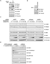
Figure 1. SGK1 expression is regulated by the Rictor pathway
A. Whole cell lysates were isolated from wild type or Rictor −/− mouse embryonic fibroblasts (MEFs) in serum starvation conditions for 24 hours. In another experimental condition, 10% FBS was added to the serum-starved cells for 1.5 hours before harvesting. Equal amounts of whole cell lysates were immunoblotted with the indicated antibodies. B. Immunoblot analysis of wild type or Rictor−/− MEFs transfected with the Myc-Rictor plasmid (with empty vector as a negative control) together with pBabe-Puro retroviral empty vector constructs. Twenty-four hours post-transfection, the cells were treated with 1 μg/ml puromycin for 48–72 hours to kill the non-transfected cells prior to collecting the whole cell lysates for immunoblots. C. HeLa cells were infected with the indicated lentiviral shRNA constructs and selected with 1μg/ml puromycin to eliminate the non-infected cells. The resulting HeLa cell lines were arrested in the M phase by incubation with nocodazole for 18 hours and then released into the G1 phase. At the indicated time points, cell lysates were collected for immunoblot analysis. D. HeLa cells were infected with lentiviral shRictor construct (with shGFP as a negative control) and selected with 1 μg/ml puromycin to eliminate the non-infected cells. The resulting cell lines were treated with 20 μM LY294002, and at the indicated time points, whole cell lysates (WCL) were collected for immunoblot analysis. (see also Figure S1)
Figure 2
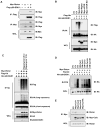
Figure 2. Rictor promotes SGK1 ubiquitination in a Cullin-1-dependent manner
A. Immunoblot (IB) analysis of whole cell lysates (WCL) and immunoprecipitates (IP) derived from 293T cells transfected with Flag-Δ60-SGK1 and Myc-Rictor constructs. Thirty hours post-transfection, cells were treated with 10 μM MG132 for 10 hours to block the proteasome pathway before harvesting. B. Immunoblot analysis of whole cell lysates (WCL) and anti-HA immunoprecipitates derived from 293T cells transfected with the indicated plasmids. Twenty hours post-transfection, cells were treated with the proteasome inhibitor MG132 overnight before harvesting. C. Immunoblot (IB) analysis of whole cell lysates (WCL) and anti-HA immunoprecipitates of 293T cells transfected with the indicated plasmids. Twenty hours post-transfection, cells were treated with the proteasome inhibitor MG132 overnight, or 100 nM Rapamycin for the indicated period of time before harvesting. D. Expression of a dominant negative form of Cullin-1 blocks the ability of Rictor to promote SGK1 ubiquitination. Immunoblot (IB) analysis of whole cell lysates (WCL) and immunoprecipitates (IP) derived from 293T cells transfected with HA-Δ60-SGK1, Myc-Rictor and His-Ub in the presence of various dominant negative forms of Cullin family members. Twenty hours post-transfection, cells were treated with the proteasome inhibitor MG132 overnight before harvesting. The whole cell lysates were collected in EDTA-free lysis buffer and the His-pull down was carried out in the presence of 8 M Urea to disrupt possible protein interactions. E. Immunoblot (IB) analysis of whole cell lysates (WCL) and immunoprecipitates (IP) derived from 293T cells transfected with the indicated Myc-Cullin constructs. Thirty hours post-transfection, cells were treated with 10 μM MG132 for 10 hours to block the proteasome pathway before harvesting. (see also Figure S2)
Figure 3
Figure 3. Rictor interacts with Cullin-1 and Rbx1 in vivo
A. Immunoblot (IB) analysis of 293T cell whole cell lysates (WCL) and anti-Cullin-1 immunoprecipitates (IP). Mouse IgG was used as a negative control for the immunoprecipitation procedure. WCL were collected with CHAPS buffer and IPs were washed with CHAPS buffer. B. Immunoblot (IB) analysis of 293T cell whole cell lysates (WCL) and anti-Rictor immunoprecipitates (IP). Rabbit IgG was used as a negative control for the immunoprecipitation procedure. WCL were collected with EBC buffer and IPs were washed with NETN buffer. C. Immunoblot (IB) analysis of 293T cell whole cell lysates (WCL) and anti-Rictor, anti-Sin1 and anti-mTOR immunoprecipitates (IP). Rabbit IgG was used as a negative control for the immunoprecipitation procedure. WCL were collected with EBC buffer and IPs were washed with NETN buffer. D. Immunoblot (IB) analysis of whole cell lysates (WCL) and immunoprecipitates (IP) derived from HeLa cells transfected with the HA-Cullin-1 construct. E. Immunoblot (IB) analysis of whole cell lysates (WCL) and immunoprecipitates (IP) derived from 293T cells transfected with HA-Rbx1 construct. Thirty hours post-transfection, cells were pretreated with 10 μM MG132 for 10 hours to block the proteasome pathway before harvesting. F. Immunoblot (IB) analysis of whole cell lysates (WCL) and immunoprecipitates (IP) derived from 293T cells transfected with Myc-Rictor together with the indicated HA-Rbx1 constructs. Thirty hours post-transfection, cells were pretreated with 10 μM MG132 for 10 hours to block the proteasome pathway before harvesting. G. Immunoblot analysis of HeLa cells transfected with the indicated siRNA oligonucleotides. The control lanes are scrambled E2F-1 siRNA and siRNA against firefly luciferase; siRNA, short interfering RNA. see also Figure S3)
Figure 4
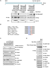
Figure 4. Rictor is phosphorylated in vivo at T1135
A. Immunoblot (IB) analysis of whole cell lysates (WCL) and immunoprecipitates (IP) derived from 293T cells transfected with the indicated Myc-Rictor constructs together with various HA-tagged AGC family of kinases. B. Sequence alignment of the putative Rictor T1135 phosphorylation site across different species. C. HeLa cells were infected with the indicated lentiviral shRNA constructs and selected with 1μg/ml puromycin to eliminate the non-infected cells. Cell lysates were collected for immunoblot analysis. D. 293T cells were transfected with the indicated Myc-tagged Rictor constructs. Thirty-six hours post-transfection, whole cell lysates were collected and the mTORC2 complex was purified by Myc-immunoprecipitation. The Myc-immunoprecipitates were incubated in vitro with the biochemically purified inactive Akt1 (from Upstate) in the presence of ATP and the kinase reaction buffer. Thirty minutes later, the reaction was stopped by the addition of the loading buffer. Akt1 phosphorylation status was examined by immunoblot analysis. (see also Figure S4)
Figure 5
Figure 5. Phosphorylation of Rictor at T1135 disrupts the interaction between Rictor and Cullin-1
A. HeLa cells were transiently transfected with HA-WT-Rictor and Myc-T1135E-Rictor constructs. Thirty hours post-transfection, whole cell lysates were collected in CHAPS buffer and subjected to gel filtration chromography. The relative band intensities for HA-WT-Rictor, Myc-T1135E-Rictor and p-T1135-Rictor at the indicated fractionations were quantitated. The original immunoblots were shown in Figure S5E. B-C. Immunoblot (IB) analysis of whole cell lysates (WCL) and immunoprecipitates (IP) derived from 293T cells transfected with the HA-Cullin-1 construct. Anti-Rictor (B) or anti-p-T1135-Rictor (C) immunoblot analysis was performed on two identical immunoprecipitations to illustrate that Cullin-1 does not interact with T1135-phosphorylated Rictor species. D. Immunoblot (IB) analysis of whole cell lysates (WCL) and immunoprecipitates (IP) derived from 293T cells transfected with the indicated Myc-Rictor and GST-Cullin-1 constructs. E. Autoradiography of 35S-labelled Cullin-1 bound to the indicated GST-fusion proteins. F. HA-Cullin-1 expressing HeLa cells transiently transfected with the Flag-14-3-3 construct. Eighteen hours post-transfection, cells were serum starved for 24 hours. After the addition of 100 nM Insulin, whole cell lysates (WCL) were collected at the indicated time points for immunoblot analysis with the indicated antibodies, for Flag-IP and GST pull-down assays to determine Rictor/14-3-3 interaction, and for HA-IP to determine Rictor/Cullin-1 interaction. G. HA-Cullin-1 expressing HeLa cells were treated with 20 μM LY294002. At the indicated time points, whole cell lysates (WCL) were collected for immunoblot analysis with the indicated antibodies, for HA-IP to determine Rictor/Cullin-1 interaction, and GST pull-down assays to determine Rictor/14-3-3 interaction. (see also Figure S5)
Figure 6
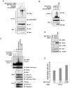
Figure 6. Phosphorylation of Rictor at T1135 reduces the ability of Rictor to ubiquitinate SGK1
A. Immunoblot (IB) analysis of whole cell lysates (WCL) and anti-HA immunoprecipitates of 293T cells transfected with the indicated plasmids. Twenty hours post-transfection, cells were treated with the proteasome inhibitor MG132 overnight before harvesting. B. Immunoblot (IB) analysis of whole cell lysates (WCL) and immunoprecipitates (IP) derived from 293T cells transfected with HA-Δ60-SGK1 together with His-Ub and various Myc-Rictor constructs. Twenty hours post-transfection, cells were treated with the proteasome inhibitor MG132 overnight before harvesting. The whole cell lysates were collected in EDTA-free lysis buffer and the His-pull down was carried out in the presence of 8 M Urea to disrupt possible protein interactions. C. Rictor/Cullin-1 promotes SGK1 ubiquitination in vitro. Immunopurified Cullin-1/Rictor complexes were incubated with purified recombinant SGK proteins (from Genway), purified E1, E2 and ubiquitin as indicated in 30°C for 45 minutes. The ubiquitination reaction products were resolved by SDS-PAGE and probed with the indicated antibodies. D-E. Wild type or Rictor−/− MEFs were transfected with the indicated Rictor plasmids (with empty vector as a negative control) together with the pBabe-Puro retroviral empty vector. Twenty-four hours post-transfection, the cells were treated with 1 μg/ml puromycin for 48–72 hours to kill the non-transfected cells prior to collecting the whole cell lysates for immunoblots (D), or subjected to BrdU analysis (E). Results are shown as means ± s.d. for three sets of experiments. (see also Figure S6)
Figure 7
Figure 7
Proposed model for the Rictor/Cullin-1 pathway to control SGK1 turnover.

References

    1. Beausoleil SA, Villen J, Gerber SA, Rush J, Gygi SP. A probability-based approach for high-throughput protein phosphorylation analysis and site localization. Nat Biotechnol. 2006;24:1285–1292. - PubMed
    1. Belova L, Sharma S, Brickley DR, Nicolarsen JR, Patterson C, Conzen SD. Ubiquitin-proteasome degradation of serum- and glucocorticoid-regulated kinase-1 (SGK-1) is mediated by the chaperone-dependent E3 ligase CHIP. Biochem J. 2006;400:235–244. - PMC - PubMed
    1. Boehm JS, Hession MT, Bulmer SE, Hahn WC. Transformation of human and murine fibroblasts without viral oncoproteins. Mol Cell Biol. 2005;25:6464–6474. - PMC - PubMed
    1. Bogusz AM, Brickley DR, Pew T, Conzen SD. A novel N-terminal hydrophobic motif mediates constitutive degradation of serum- and glucocorticoid-induced kinase-1 by the ubiquitin-proteasome pathway. Febs J. 2006;273:2913–2928. - PubMed
    1. Brickley DR, Mikosz CA, Hagan CR, Conzen SD. Ubiquitin modification of serum and glucocorticoid-induced protein kinase-1 (SGK-1) J Biol Chem. 2002;277:43064–43070. - PubMed

Publication types

MeSH terms