. 2010 Oct 8;401(1):58-63.
doi: 10.1016/j.bbrc.2010.09.008. Epub 2010 Sep 15.

Solution NMR structure of the V27A drug resistant mutant of influenza A M2 channel

Affiliations

Solution NMR structure of the V27A drug resistant mutant of influenza A M2 channel

Rafal M Pielak et al. Biochem Biophys Res Commun. .

Abstract

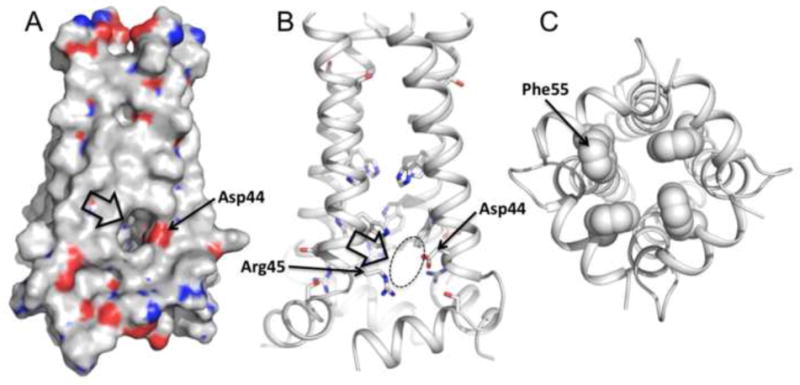
The M2 protein of influenza A virus forms a proton-selective channel that is required for viral replication. It is the target of the anti-influenza drugs, amantadine and rimantadine. Widespread drug resistant mutants, however, has greatly compromised the effectiveness of these drugs. Here, we report the solution NMR structure of the highly pathogenic, drug resistant mutant V27A. The structure reveals subtle structural differences from wildtype that maybe linked to drug resistance. The V27A mutation significantly decreases hydrophobic packing between the N-terminal ends of the transmembrane helices, which explains the looser, more dynamic tetrameric assembly. The weakened channel assembly can resist drug binding either by destabilizing the rimantadine-binding pocket at Asp44, in the case of the allosteric inhibition model, or by reducing hydrophobic contacts with amantadine in the pore, in the case of the pore-blocking model. Moreover, the V27A structure shows a substantially increased channel opening at the N-terminal end, which may explain the faster proton conduction observed for this mutant. Furthermore, due to the high quality NMR data recorded for the V27A mutant, we were able to determine the structured region connecting the channel domain to the C-terminal amphipathic helices that was not determined in the wildtype structure. The new structural data show that the amphipathic helices are packed much more closely to the channel domain and provide new insights into the proton transfer pathway.


Figures

Figure 1
Figure 1
Amide and methyl spectra of the V27A18-60 mutant display residue specific assignment of backbone amine and sidechain methyl groups, respectively. a. 1H-15N tr-HSQC spectrum of 15N-, 13C, and 85% 2H-labeled protein. b. 1H-13C HSQC spectrum of methyl groups of uniformly 15N-, 13C-labeled protein. Both spectra were recorded at 30°C, pH 7.5, and 1H frequency of 600MHz.
Figure 2
Figure 2. Structures of WT and the V27A mutant
a. Superposition of 15 low energy structures of WT18-60 (2RLF)[16] and b. V27A18-60 (2KWX). The V27A structures were calculated using restraints summarized in Table 1. c. Ribbon representation of the WT structure (2RLF) and d. the V27A structure (2KWX). Compared to WT, the structure ensemble of V27A shows better-defined arrangement of the AP helices relative to the pore domain due to more long-range NOEs.
Figure 3
Figure 3
The size of the channel N-terminal entrance of a. the V27A mutant (2KWX), b. WT (2RLF), and c. the S31N mutant (2KIH) indicated by double headed arrow. The side chains at residue position 27 constrict the channel entrance. Mutating valine at this position to alanine doubles the diameter of the channel opening (∼2.5 Å in WT and S31N mutant, ∼5 Å in the V27A mutant). d,e,f The pore surfaces calculated using the program HOLE. d. The WT structure displays two constrictions in the N terminus at the positions 27 and 30. e. The V27A mutant displays one constriction at position 30. f. The S31N mutant is constricted at the position 27, but due to the serine to asparagine substitution at the position 31 the channel forms looser tetramer that result in somewhat larger diameter around Ser30. All of the structures have their C-termini tightly constricted to ∼1.5 Å by side chains of His37 and Trp41.
Figure 4
Figure 4
The proton exit suggested by the structure. a. Van der Waals surface and b. ribbon representations of the V27A structure show a lateral opening around Asp44 and Arg45. Asp44 is positioned near the lipid headgroups of presumed bilayer where it is accessible by water molecules, thus is capable of mediating C-terminal proton or hydronium exit. c. The C-terminal base of the channel is sealed by the hydrophobic side chains of Phe55, making it impermeable to either water molecules or protons.

Similar articles

Cited by

References

    1. Hay AJ, Wolstenholme AJ, Skehel JJ, Smith MH. The molecular basis of the specific anti-influenza action of amantadine. Embo J. 1985;4:3021–4. - PMC - PubMed
    1. Helenius A. Unpacking the incoming influenza virus. Cell. 1992;69:577–578. - PubMed
    1. Pinto LH, Holsinger LJ, Lamb RA. Influenza virus M2 protein has ion channel activity. Cell. 1992;69:517–28. - PubMed
    1. Davies WL, Grunert RR, Haff RF, McGahen JW, Neumayer EM, Paulshock M, Watts JC, Wood TR, Hermann EC, Hoffmann CE. Antiviral Activity of 1-Adamantanamine (Amantadine) Science. 1964;144:862–3. - PubMed
    1. Bright RA, Shay DK, Shu B, Cox NJ, Klimov AI. Adamantane resistance among influenza A viruses isolated early during the 2005-2006 influenza season in the United States. Jama. 2006;295:891–4. - PubMed

Publication types

LinkOut - more resources