. 2011 Jan 1;81(1):24-31.
doi: 10.1016/j.bcp.2010.09.001. Epub 2010 Sep 15.

Organ-specific carboxylesterase profiling identifies the small intestine and kidney as major contributors of activation of the anticancer prodrug CPT-11

Affiliations

Organ-specific carboxylesterase profiling identifies the small intestine and kidney as major contributors of activation of the anticancer prodrug CPT-11

M Jason Hatfield et al. Biochem Pharmacol. .

Abstract

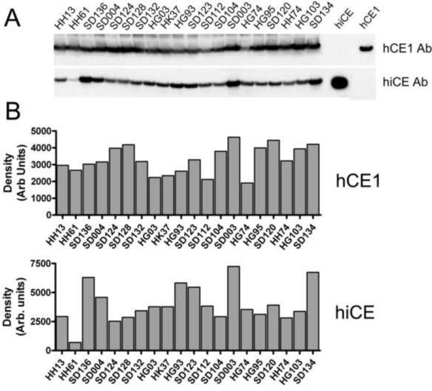
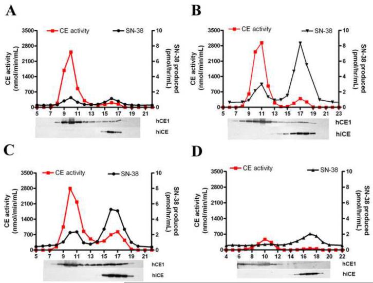
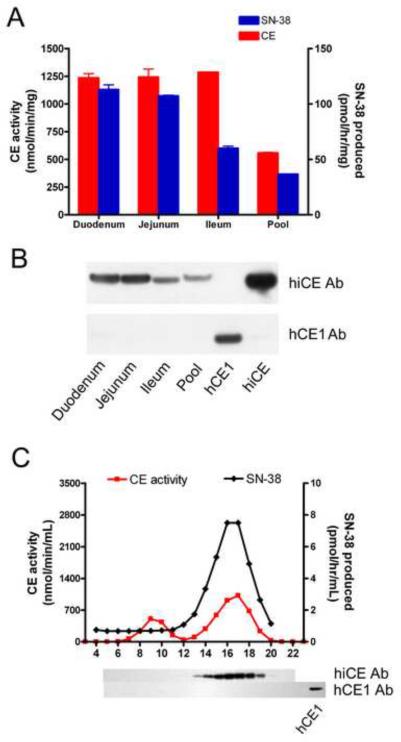
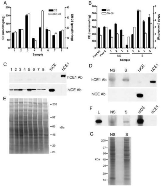
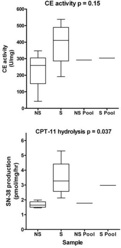
The activation of the anticancer prodrug CPT-11, to its active metabolite SN-38, is primarily mediated by carboxylesterases (CE). In humans, three CEs have been identified, of which human liver CE (hCE1; CES1) and human intestinal CE (hiCE; CES2) demonstrate significant ability to hydrolyze the drug. However, while the kinetic parameters of CPT-11 hydrolysis have been measured, the actual contribution of each enzyme to activate the drug in biological samples has not been addressed. Hence, we have used a combination of specific CE inhibition and conventional chromatographic techniques to determine the amounts, and hydrolytic activity, of CEs present within human liver, kidney, intestinal and lung specimens. These studies confirm that hiCE demonstrates the most efficient kinetic parameters for CPT-11 activation, however, due to the high levels of hCE1 that are expressed in liver, the latter enzyme can contribute up to 50% of the total of drug hydrolysis in this tissue. Conversely, in human duodenum, jejunum, ileum and kidney, where hCE1 expression is very low, greater than 99% of the conversion of CPT-11 to SN-38 was mediated by hiCE. Furthermore, analysis of lung microsomal extracts indicated that CPT-11 activation was more proficient in samples obtained from smokers. Overall, our studies demonstrate that hCE1 plays a significant role in CPT-11 hydrolysis even though it is up to 100-fold less efficient at drug activation than hiCE, and that drug activation in the intestine and kidney are likely major contributors to SN-38 production in vivo.


Figures

Figure 1
Figure 1
Analysis of CE expression in human liver microsomes A – Western analysis of hCE1 and hiCE expression in human liver microsomal extracts. Purified proteins (50ng of each) were used as positive controls and are indicated on the right hand side of the image. B – Densitometric quantitation of the western analyses depicted in panel A.
Figure 2
Figure 2
Chromatographic separation of CEs expressed in human liver microsomes. Four individual microsomal extracts (HH61, SD136, SD132 and HH13) were run on Sephacryl S-200HR and fractions were assayed for both CE activity using o-NPA and CPT-11 as substrates, and immuno-reactive protein. Each panel represents the elution profile for a different extract, with expression levels ranging from high hCE1, low hiCE (HH61; panel A); moderate hCE1, high hiCE (SD136; panel B); moderate hCE1, moderate hiCE (SD132; panel C); to low hCE1, moderate hiCE (HH13; panel D). CE activity and CPT-11 hydrolysis are indicated by the red and the black lines, respectively. Western analysis of the individual fractions, designed to identify hCE1 and hiCE, are aligned and indicated below each graph.
Figure 3
Figure 3
Analysis of CE expression in human intestinal microsomes. A – CE and CPT-11 hydrolytic activities of microsomal extracts isolated from different regions of the small intestine. The duodenum, jejunum and ileum samples are isolated from the same individual, and the extract labeled `Pool' represents small intestinal microsomes mixed from 8 donors. B – Western analysis of the samples described in A. Samples were probed with either an antibody recognizing hiCE (upper panel) or hCE1 (lower panel). As positive controls for these experiments, 50ng of purified proteins were included on the membrane. C – Chromatographic profile of the CEs present within the duodenum microsomal extract. The extract was separated on Sephacryl S-200 and fractions were assayed in an identical fashion to that described in the legend for Figure 2. Western analyses for hiCE and hCE1 are indicated under the respective fractions. A positive control is included for hCE1 to confirm reactivity of the antibody.
Figure 4
Figure 4
Analysis of CE expression in human lung and kidney microsomes. A - Graph indicating the levels of CE activity (black bars) and SN-38 production (white bars) from individual kidney microsomal samples. B – Graph indicating the levels of CE activity (black bars) and SN-38 production (white bars) by both pooled and individual lung microsomal samples. NS – non smokers; S – smokers. C – Western analysis of hiCE and hCE1 expression in individual human kidney microsomal extracts. Purified hiCE and hCE1 protein (50ng) were included as positive controls. D – Western analysis of hiCE and hCE1 expression in pooled human lung microsomal extracts isolated from 4 individuals. Purified hiCE and hCE1 protein (50ng) were included as positive controls. As before, NS – non smokers; S – smokers. E – Coomassie blue stained gel demonstrating equal loading of the samples analyzed in panel C. F – Prolonged exposure of a western analysis using larger amounts (200μg) of lung microsomal extracts following immunoblotting with hiCE antibody. L - human liver microsomes. G – Coomassie blue stained gel demonstrating equal loading of the samples analyzed in the western analyses. As above, NS – non smokers; S – smokers.
Figure 5
Figure 5
CE expression and CPT-11-converting activities of human lung microsomes. Graphs comparing the levels of CE activity (upper panel) and CPT-11 hydrolysis (lower panel) in microsomal extracts isolated from lung tissue of both smokers (S) and non-smokers (NS). Data for individual samples were analyzed using the unpaired T-test, with p values for each dataset indicated on the graph. Samples marked as `Pool' represent mixed extracts obtained from 4 individuals.

References

    1. Tanizawa A, Fujimori A, Fujimori Y, Pommier Y. Comparison of topoisomerase I inhibition, DNA damage, and cytotoxicity of camptothecin derivatives presently in clinical trials. J Natl Cancer Inst. 1994;86:836–42. - PubMed
    1. Danks MK, Morton CL, Krull EJ, Cheshire PJ, Richmond LB, Naeve CW, et al. Comparison of activation of CPT-11 by rabbit and human carboxylesterases for use in enzyme/prodrug therapy. Clin Cancer Res. 1999;5:917–24. - PubMed
    1. Danks MK, Morton CL, Pawlik CA, Potter PM. Overexpression of a rabbit liver carboxylesterase sensitizes human tumor cells to CPT-11. Cancer Res. 1998;58:20–2. - PubMed
    1. Humerickhouse R, Lohrbach K, Li L, Bosron W, Dolan M. Characterization of CPT-11 hydrolysis by human liver carboxylesterase isoforms hCE-1 and hCE-2. Cancer Res. 2000;60:1189–92. - PubMed
    1. Khanna R, Morton CL, Danks MK, Potter PM. Proficient metabolism of CPT-11 by a human intestinal carboxylesterase. Cancer Res. 2000;60:4725–8. - PubMed

Publication types

MeSH terms