. 2010 Dec 15;171(3):893-909.
doi: 10.1016/j.neuroscience.2010.08.059. Epub 2010 Sep 15.

Ovarian steroid regulation of the midbrain corticotropin releasing factor and urocortin systems in macaques

Affiliations

Ovarian steroid regulation of the midbrain corticotropin releasing factor and urocortin systems in macaques

R L Sanchez et al. Neuroscience. .

Abstract

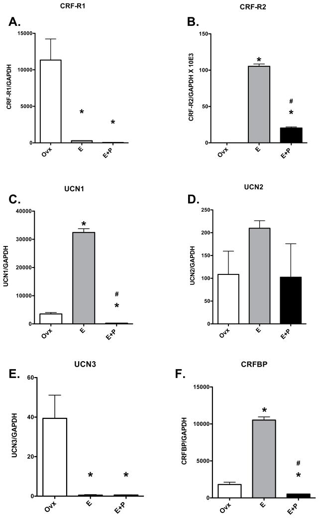
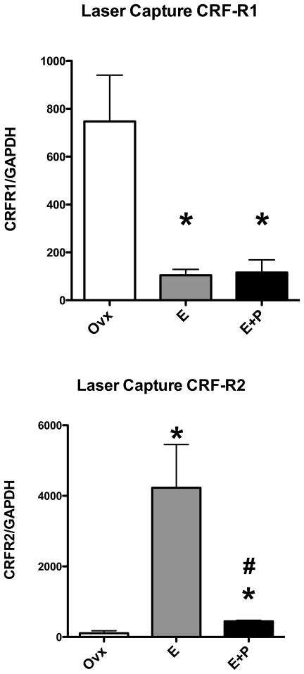
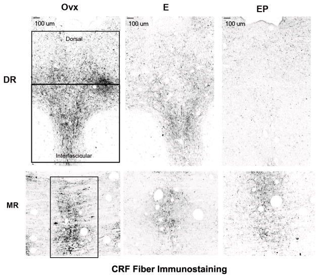
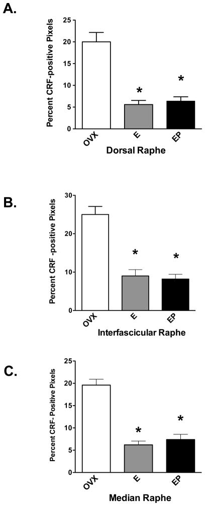
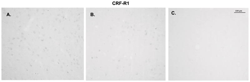
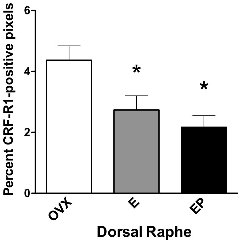
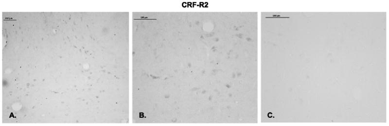
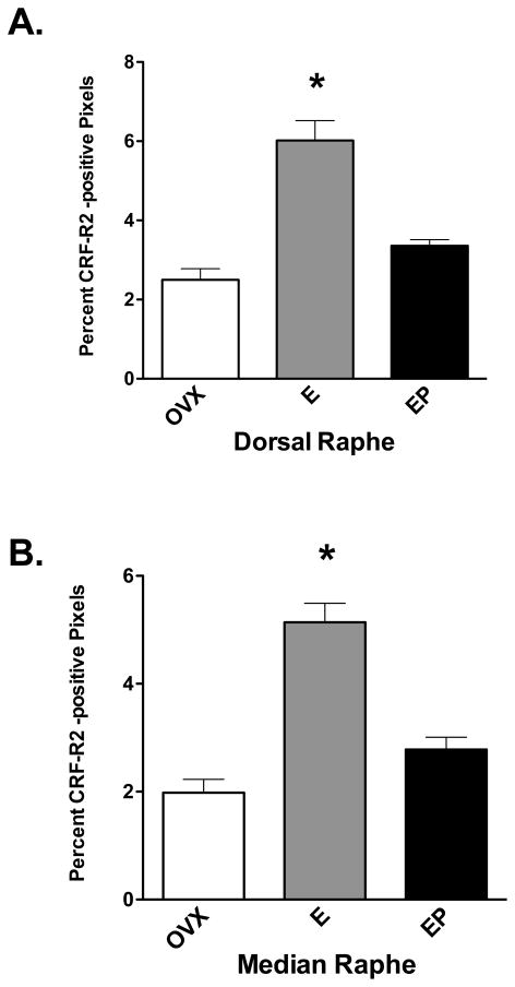
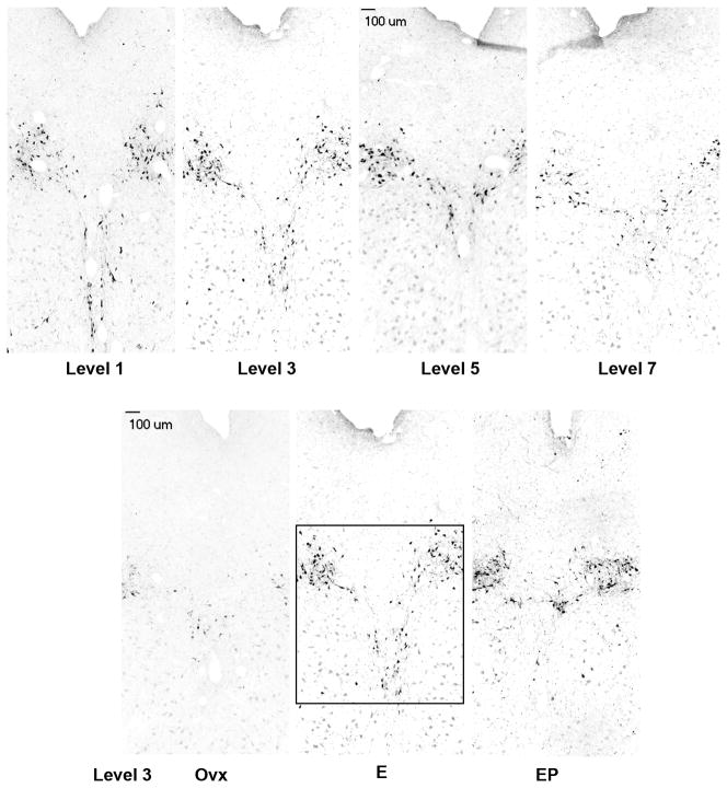
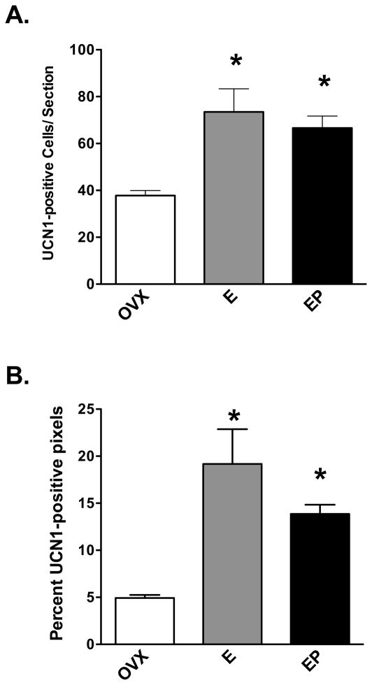
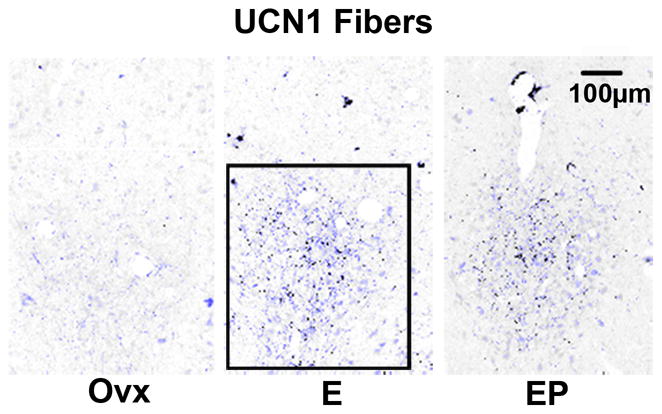
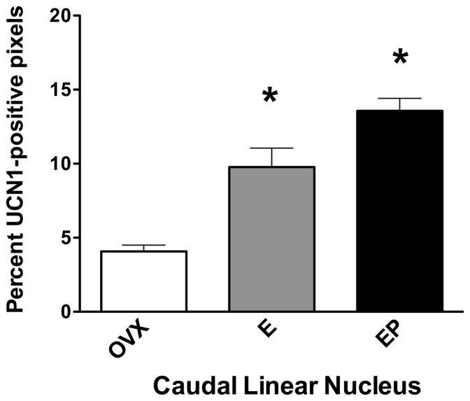
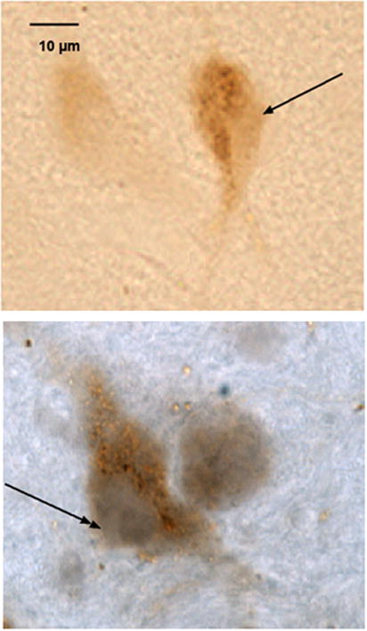
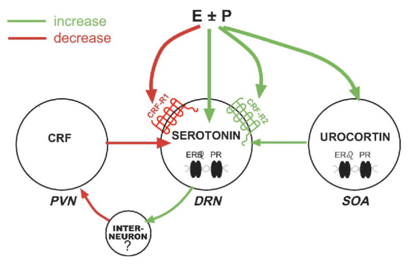
A significant number of postmenopausal women report increased anxiety and vulnerability to stress, which has been linked to decreased secretion of ovarian steroids. Communication between the serotonin system and the corticotropin releasing factor (CRF) system determines stress sensitivity or resilience. This study examines the effects of the ovarian steroids, estradiol (E) and progesterone (P) on the CRF system components that impact serotonin neurons in the midbrain of nonhuman primates. Ovariectomized rhesus macaques were treated with placebo, E alone for 1 month, or E supplemented with P for the last 2 weeks. Quantitative (q)RT-PCR and immunocytochemistry were employed. E±P treatment decreased CRF-R1 and increased CRF-R2 gene expression in hemi-midbrain blocks and in laser captured serotonin neurons. Also in hemi-midbrains, E treatment increased urocortin 1 (UCN1) and CRFBP gene expression, but supplemental P treatment reversed these effects. E±P decreased CRF fiber density in the dorsal, interfascicular and median raphe nuclei and decreased CRF-R1 immunostaining in the dorsal raphe. E increased CRF-R2 immunostaining in the dorsal and median raphe. E±P increased UCN1 immunostaining in the cell bodies and increased UCN1 fiber density in the caudal linear nucleus. Estrogen receptor beta (ERβ), but not ERα was detected in the nucleus of UCN1-positive neurons. While the mechanism of ovarian hormone regulation of the midbrain CRF system requires further investigation, these studies clearly demonstrate another pathway by which ovarian hormones may have positive effects on anxiety and mood regulation.


Figures

Figure 1
Figure 1
An Illustration of the dissection of the midbrain and preparation of tissue for assay. In panel A, the midbrain and brainstem were removed from the cerebellum and bissected along the saggital plane. One half was immersion fixed in RNA later and then frozen at −80C until RNA extraction for qRT-PCR employing a Taqman Custom Expression Array. In panel B, the midbrain was removed from the hypothalamus with a rostral coronal cut and from the brainstem and cerebellum with a caudal coronal cut. In animals that were perfused with RNAlater, this block of tissue was sectioned coronally at 7 μm for laser capture. In animals that were perfused with 4% formaldehyde this block of tissue was sectioned coronally at 25 μm for ICC.
Figure 2
Figure 2
Relative expression of stress-related genes in a hemi-midbrain block from ovariectomized monkeys treated with placebo (Ovx), estrogen (E) or estrogen +progesterone (E+P) for one month (n=4/group). Gene expression was determined with custom Taqman expression cards from ABI. Each transcript is normalized to GAPDH. A. The relative expression of CRF-R1 was significantly decreased by E and E+P treatments compared to the Ovx control group (ANOVA p < 0.0014). B. The relative expression of CRF-R2 was significantly increased by E treatment compared to the Ovx control group and then significantly reduced by P treatment. However, expression in the P group remained significantly higher than in the Ovx control group (ANOVA p < 0.0001). C. The relative expression of UCN1 was significantly increased by E treatment compared to the Ovx control group and then significantly reduced by P treatment (ANOVA p < 0.0001). D. The relative expression of UCN2 was not affected by ovarian steroids. E. The relative expression of UCN3 was significantly reduced by E and E+P treatment compared to the Ovx control group (ANOVA p < 0.0029). However, the expression was at the limit of detection of the assay. F. The relative expression of CRFBP was significantly increased by E treatment compared to the Ovx control group and then suppressed by supplemental P treatment (ANOVA p < 0.0001). Asterisks (*) mean p < 0.05 compared to Ovx control group and crosshairs (#) mean p < 0.05 compared to E group, Student-Newman-Keuls posthoc pairwise analysis.
Figure 3
Figure 3
Relative expression of CRF-R1 and CRF-R2 in laser captured serotonin neurons from ovariectomized monkeys treated with placebo (Ovx), estrogen (E) or estrogen +progesterone (E+P) for one month (n=3/group). Gene expression was determined with custom Taqman expression cards from ABI after amplification with a mix of primers. Each transcript is normalized to GAPDH. Top. The relative expression of CRF-R1 was significantly decreased by E and E+P treatments compared to the Ovx control group (ANOVA p < 0.0124). Bottom. The relative expression of CRF-R2 was significantly increased by E treatment compared to the Ovx control group and then significantly reduced by supplemental P treatment (ANOVA p < 0.0044). Symbols are as described in Figure 2.
Figure 4
Figure 4
Representative stereological montages of CRF-positive fibers in the dorsal (DR) and median (MR) raphe nuclei from ovariectomized monkeys treated with placebo (Ovx), estrogen (E) or estrogen +progesterone (E+P) for one month (n=5/group). The boxes demarcate the regions of interest of the raphe that were analyzed at this level. In the dorsal raphe (DR), the top area was designated the dorsal aspect of the dorsal raphe nucleus and the lower area was designated the interfascicular dorsal raphe nucleus. The size of the region of interest varied at each anatomical level, but it was always held constant across all animals. There is an apparent decrease in CRF-positive fibers in the animals treated with E or E+P compared to the Ovx control group.
Figure 5
Figure 5
Histograms representing the percent of CRF-positive pixels within the dorsal, interfascicular and median raphe nuclei from ovariectomized monkeys treated with placebo (Ovx), estrogen (E) or estrogen + progesterone (E+P) for one month (n=5/group). A. E and E+P significantly decreased the percent of CRF-positive pixels in the dorsal segment of the dorsal raphe nucleus in all four levels analyzed (ANOVAs range p < 0.0001 to 0.0006; not shown). When all levels were combined, E and E+P significantly decreased the mean percent of CRF-positive pixels compared to Ovx placebo-treated controls (ANOVA, p<0.0001). B. E and E+P significantly decreased the percent of CRF-positive pixels in the interfascicular segment of the dorsal raphe nucleus in all four levels analyzed (ANOVAs range p < 0.0001 to 0.02; not shown). When the interfascicular levels of the dorsal raphe nucleus were combined, E and E+P significantly decreased the mean percent of CRF-positive pixels compared to Ovx placebo-treated controls (ANOVA, p<0.0001). C. E and E+P significantly decreased the percent of CRF-positive pixels in the median raphe in all four levels analyzed (ANOVAs range p < 0.0001 to 0.02; not shown). When the levels were combined, E and E+P significantly decreased the percent of CRF-positive pixels compared to Ovx placebo-treated controls (ANOVA, p<0.05). Symbols are as described in Figure 2.
Figure 6
Figure 6
Immunostaining of CRF-R1 in the dorsal raphe nucleus. A. Dorsal raphe nucleus from Ovx control animal immunostained with the CRF-R1 antibody diluted 1/500 in buffer. B. Dorsal raphe nucleus from Ovx control animal immunostained with the CRF-R1 antibody diluted 1/2000 in buffer. This was the highest dilution to produce immunostaining. C. Raphe section from the same animal incubated with the 1/2000 dilution of the primary antibody preabsorbed with 500 μg CRF-R1 peptide for 24 hours before incubation with the tissue section.
Figure 7
Figure 7
Histogram representing the percent of CRF-R1-positive pixels within the dorsal raphe nucleus from ovariectomized monkeys treated with placebo (Ovx), estrogen (E) or estrogen +progesterone (E+P) for one month (n=3/group). A total of 5 levels at 500 μm intervals were examined. E and E + P significantly decreased the percent of CRF-R1-positive pixels in the third level of the dorsal raphe nucleus (ANOVA, p <0.0025; not shown). When all levels were combined, E and E+P significantly decreased the mean percent of CRF-R1-positive pixels compared to OVX placebo-treated controls (ANOVA, p<0.03). Symbols are as described in Figure 2.
Figure 8
Figure 8
Immunostaining for CRF-R2 in the ventral part of the dorsal raphe nucleus. A. Low power magnification of CRF-R2 immunostaining with a 1/750 dilution of the antibody. B. High magnification of CRF-R2 immunostaining with a 1/750 dilution of the antibody. C. Raphe section from the same animal incubated with 1/750 dilution of the primary antibody preabsorbed with 1 mg CRF-R2 peptide for 24 hours before incubation with the tissue section.
Figure 9
Figure 9
Histograms representing the percent of CRF-R2-positive pixels in the dorsal and median raphe nuclei from ovariectomized monkeys treated with placebo (Ovx), estrogen (E) or estrogen +progesterone (E+P) for one month (n=5/group). A. The E-treated group showed a significantly higher percent of CRF-R2-positive pixels in the dorsal raphe nucleus compared to Ovx placebo- or E+P-treated groups in 4 of 5 levels examined (ANOVAs range p < 0.0001 to 0.014; not shown). When all 5 levels were combined, E and E+P significantly increased the mean percent of CRF-R2-positive pixels compared to Ovx placebo-treated controls (ANOVA, p<0.05). B. The E-treated group showed a significantly higher percent of CRF-R2-positive pixels in the median raphe nucleus compared to Ovx placebo- or E+P-treated groups at all 3 levels examined (ANOVAs range p < 0.0001 to 0.018; not shown). When all 3 levels were combined, E treatment significantly increased the mean percent of CRF-R2-positive pixels compared to Ovx placebo-treated controls (ANOVA, p<0.0001). Symbols are as described in Figure 2.
Figure 10
Figure 10
Montages of UCN1 immunostained neurons in the supraoculomotor area (SOA) adjacent to the Edinger Westfal nucleus. Top panels. UCN1 immunostaining in representative sections of the SOA at 4 of the 8 levels that were subjected to quantitative analysis. Each level is 500 μm apart from a rostral to caudal direction. Bottom panels. UCN1 immunostaining in representative sections of the SOA from an ovx control, an E-treated and an E+P treated animal. There appears to be an increase in UCN1 cell staining with E and E+P treatment. The box demarcates the region of interest that was analyzed at level 3. The size of the region of interest varied at each anatomical level, but it was always held constant across all animals.
Figure 11
Figure 11
Histograms representing the number of UCN1-positive neurons and percent UCN1-positive pixels within the supraoculomotor region in each treatment group (n=5/group). A. E or E+P significantly increased the number of UCN1-positive neurons in 7 of 8 levels analyzed (ANOVAs range p < 0.0025 to 0.038; not shown). When all 8 levels were combined, E and E+P significantly increased the mean number of UCN1-positive neurons per section compared to placebo-treated controls (ANOVA, p<0.0029). B. E and E+P also significantly increased the percent of UCN1-positive pixels in the supraoculomotor region in all 8 levels analyzed (ANOVAs range p < 0.0001 to 0.037; not shown). When the levels were combined, E and E+P significantly increased the mean percent of UCN1-positive pixels compared to placebo controls (ANOVA, p<0.0001). Symbols are as described in Figure 2.
Figure 12
Figure 12
Representative montages illustrating immunostaining for UCN1 fibers in the caudal linear nucleus from ovariectomized monkeys treated with placebo (Ovx), estrogen (E) or estrogen +progesterone (E+P) for one month (n=5/group). The UCN1 immunostained fibers have been segmented from background in blue. The box demarcates the region of interest that was analyzed at this level. The size of the region of interest varied at each anatomical level, but it was always held constant across all animals.
Figure 13
Figure 13
Histogram representing the percent of UCN1-positive pixels in the caudal linear nucleus from ovariectomized monkeys treated with placebo (Ovx), estrogen (E) or estrogen +progesterone (E+P) for one month (n=5/group). E or E+P significantly increased the percent of CRF-positive pixels in the caudal linear raphe nucleus at all 3 levels examined (ANOVAs range p < 0.0034 to 0.0097; not shown). When all 3 levels were combined, E and E+P significantly increased the percent of UCN1-positive pixels compared to Ovx placebo-treated controls (ANOVA, p<0.0001). Symbols are as described in Figure 2.
Figure 14
Figure 14
Neurons in the supraoculomotor region of an E+P treated animal immunostained with antibodies against human UCN1 and nuclear estrogen receptor beta (ERβ). Top. Neuron immunostained for UCN1 only, visualized as a reddish-brown precipitate in the cytoplasm. There is no staining in the nucleus (single head arrow). Bottom. Neuron immunostained for UCN1 and ERβ. The immunostaining for ERβ appears purple and fills the nucleus (double headed arrow), while the reddish-brown precipitate for UCN1 fills the neuronal cytoplasm.
Figure 15
Figure 15
A diagram of the hypothesized interactions of ovarian hormones and midbrain stress systems. We show herein that E±P decrease CRF innervation of the dorsal and median raphe nuclei, the location of serotonin cell bodies that project to the forebrain. In addition, E±P decrease CRF-R1 and increase CRF-R2 on serotonin neurons. We also show that E±P increases UCN1 production and fiber density rostral to the dorsal raphe, and those UCN1 neurons contain ERβ. PR is also likely present in UCN1 neurons that colocalize CART (Lima et al., 2008). We have previously shown that serotonin neurons contain ERβ and PR, which in turn increase production of serotonin (Bethea, 1993, 1994, Gundlah et al., 2001). We speculate that serotonin projects to the hypothalamus and stimulates an inhibitory interneuron that inhibits CRF production in the PVN (Bethea and Centeno, 2008). This does not rule out a direct action of E±P on CRF in the PVN, but an inhibitory effect has not been observed in molecular studies.

Similar articles

Cited by

References

    1. Amat J, Tamblyn JP, Paul ED, Bland ST, Amat P, Foster AC, Watkins LR, Maier SF. Microinjection of urocortin 2 into the dorsal raphe nucleus activates serotonergic neurons and increases extracellular serotonin in the basolateral amygdala. Neuroscience. 2004;129:509–519. - PubMed
    1. Arango V, Underwood MD, Mann JJ. Serotonin brain circuits involved in major depression and suicide. Prog Brain Res. 2002;136:443–453. - PubMed
    1. Austin MC, Rice PM, Mann JJ, Arango V. Localization of corticotropin-releasing hormone in the human locus coeruleus and pedunculopontine tegmental nucleus: an immunocytochemical and in situ hybridization study. Neuroscience. 1995;64:713–727. - PubMed
    1. Bao AM, Hestiantoro A, Van Someren EJ, Swaab DF, Zhou JN. Colocalization of corticotropin-releasing hormone and oestrogen receptor-alpha in the paraventricular nucleus of the hypothalamus in mood disorders. Brain. 2005;128:1301–1313. - PubMed
    1. Barbaccia ML, Serra M, Purdy RH, Biggio G. Stress and neuroactive steroids. Int Rev Neurobiol. 2001;46:243–272. - PubMed

Publication types

MeSH terms