

Developmental changes in brain connectivity assessed using the sleep EEG
- PMID: 20833232
- PMCID: PMC4119998
- DOI: 10.1016/j.neuroscience.2010.08.071
Developmental changes in brain connectivity assessed using the sleep EEG
Abstract
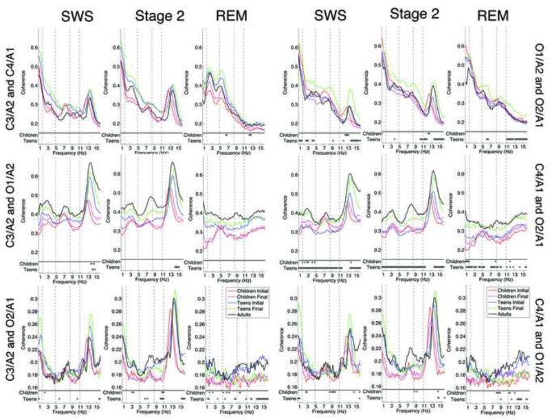
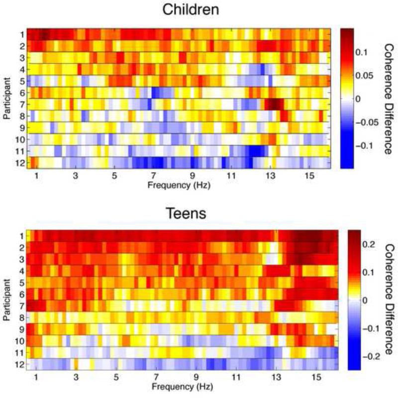
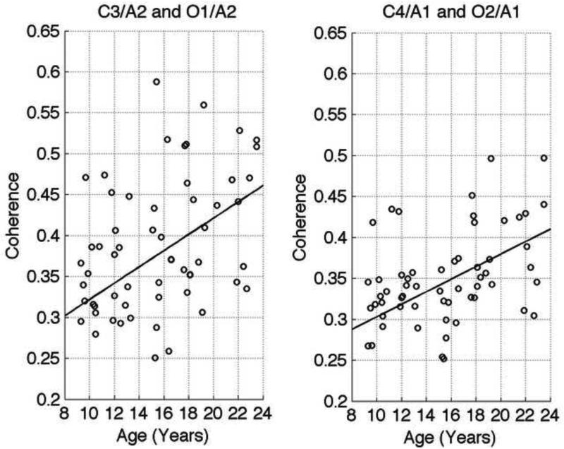
Adolescence represents a time of significant cortical restructuring. Current theories posit that during this period connections between frequently utilized neural networks are strengthened while underutilized synaptic connections are discarded. The aim of the present study was to examine the developmental evolution of connectivity between brain regions using the sleep EEG. All-night sleep EEG recordings in two longitudinal cohorts (children and teens) followed at 1.5-3 year intervals and one cross-sectional cohort (adults) were analyzed. The children and teen cohorts were 9/10 and 15/16 years at the initial assessment; ages of the adults were 20 to 23 years. Intrahemispheric, interhemispheric, and diagonal coherence was measured between all six possible pairings of two central (C3/A2 and C4/A1) and two occipital (O2/A1 and O1/A2) derivations during slow wave, stage 2, and, REM sleep. Within-subjects analyses were performed for the children and teen cohorts, and a linear regression analysis was performed across every assessment of all cohorts. Within-subject analyses revealed a maturational increase in coherence for both age cohorts, though the frequencies, sleep states, and regions differed between cohorts. Regression analysis across all age cohorts showed an overall linear increase in left and right intrahemispheric coherence for all sleep states across frequencies. Furthermore, coherence between diagonal electrode pairs also increased in a linear manner for stage 2 and REM sleep. No age-related trend was found in interhemispheric coherence. Our results indicate that sleep EEG coherence increases with age and that these increases are confined to specific brain regions. This analysis highlights the utility of the sleep EEG to measure developmental changes in brain maturation.
Copyright © 2010 IBRO. Published by Elsevier Ltd. All rights reserved.
Figures
References
-
- Achermann P, Borbely AA. Coherence analysis of the human sleep electroencephalogram. Neuroscience. 1998;85:1195–1208. - PubMed
-
- Anokhin AP, Lutzenberger W, Birbaumer N. Spatiotemporal organization of brain dynamics and intelligence: an EEG study in adolescents. Int J Psychophysiol. 1999;33:259–273. - PubMed
-
- Armitage P, Parmar M. Some approaches to the problem of multiplicity in clinical trials.. Proceedings of the XIIthe International Biometrics Conference..1986.
-
- Armitage R, Hoffmann R, Emslie G, Rintelmann J, Robert J. Sleep microarchitecture in childhood and adolescent depression: temporal coherence. Clin EEG Neurosci. 2006;37:1–9. - PubMed
-
- Ashtari M, Cervellione KL, Hasan KM, Wu J, McIlree C, Kester H, Ardekani BA, Roofeh D, Szeszko PR, Kumra S. White matter development during late adolescence in healthy males: a cross-sectional diffusion tensor imaging study. NeuroImage. 2007;35:501–510. - PubMed
Publication types
MeSH terms
Grants and funding
LinkOut - more resources
Full Text Sources
Miscellaneous
