Review
. 2011 May;29(3):311-24.
doi: 10.1016/j.ijdevneu.2010.08.007. Epub 2010 Sep 15.

Mitochondrial dysfunction and pathology in bipolar disorder and schizophrenia

Affiliations
Review

Mitochondrial dysfunction and pathology in bipolar disorder and schizophrenia

Hayley B Clay et al. Int J Dev Neurosci. 2011 May.

Abstract

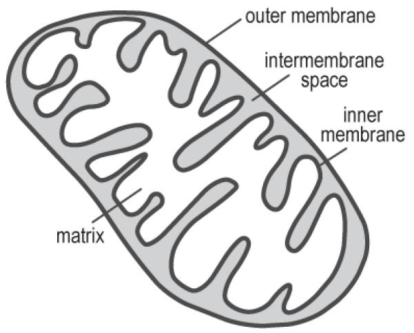
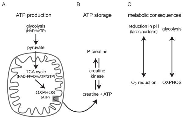
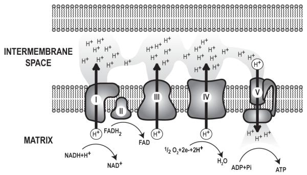
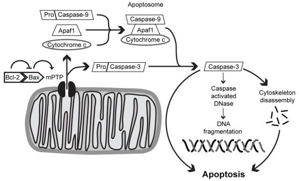
Bipolar disorder (BPD) and schizophrenia (SZ) are severe psychiatric illnesses with a combined prevalence of 4%. A disturbance of energy metabolism is frequently observed in these disorders. Several pieces of evidence point to an underlying dysfunction of mitochondria: (i) decreased mitochondrial respiration; (ii) changes in mitochondrial morphology; (iii) increases in mitochondrial DNA (mtDNA) polymorphisms and in levels of mtDNA mutations; (iv) downregulation of nuclear mRNA molecules and proteins involved in mitochondrial respiration; (v) decreased high-energy phosphates and decreased pH in the brain; and (vi) psychotic and affective symptoms, and cognitive decline in mitochondrial disorders. Furthermore, transgenic mice with mutated mitochondrial DNA polymerase show mood disorder-like phenotypes. In this review, we will discuss the genetic and physiological components of mitochondria and the evidence for mitochondrial abnormalities in BPD and SZ. We will furthermore describe the role of mitochondria during brain development and the effect of current drugs for mental illness on mitochondrial function. Understanding the role of mitochondria, both developmentally as well as in the ailing brain, is of critical importance to elucidate pathophysiological mechanisms in psychiatric disorders.


Figures

Figure 1
Figure 1
Mitochondrial anatomy: Mitochondria are intracellular organelles that contain both outer and inner membranes separated by an intermembrane space. The innermost mitochondrial compartment, the matrix, contains the enzymes necessary for tricarboxylic acid (TCA) cycle, while the inner membrane contains the components of the electron transport chain.
Figure 2
Figure 2
Energy metabolism and metabolic consequences: A) Glycolysis, a cytosolic process, breaks down glucose to pyruvate. The net yield of glycolysis is two molecules of ATP and two molecules of the electron donor NADH, which are shuttled into the mitochondria. Likewise, pyruvate crosses into the mitochondria and is converted acetyl-CoA, which enters the tricarboxylic acid (TCA) cycle. The conversion to acetyl-CoA produces NADH. The TCA cycle, then produces three molecules of NADH, one molecule of the electron donor FADH2, as well as one molecule of ATP or GTP. NADH and FADH2 donate electrons to the electron transport chain of the inner mitochondrial membrane, where ATP is synthesized during oxidative phosphorylation (OXPHOS). Each glucose molecule produces 36 ATP molecules when metabolized by OXPHOS. B) Creatine kinase uses ATP to phosphorylate creatine and produce phosphocreatine (P-creatine), a stable product for extended storage of ATP. During periods of high energy demand, creatine kinase acts as a phosphatase to retrieve ATP from P-creatine. C) metabolic shift from OXPHOS to glycolysis results in cellular acidification through lactic acidosis, a metabolization from pyruvate to lactate. Conversely, OXPHOS activity measured as reduction in O2.
Figure 3
Figure 3
The electron transport chain: Electron transport chain complexes I-V are part of the inner mitochondrial membrane. NADH and FADH2 from the tricarboxylic acid (TCA) cycle donate electrons to complexes I and II, respectively. These electrons are transferred to complex III and complex IV, sequentially. With each transfer, the electrons release energy. Energy release from electrons is coupled to the movement of protons from the matrix to the intermembrane space, resulting in a proton gradient across the inner mitochondrial membrane. Upon reaching complex IV, electron pairs combine with ½ O2 and 2H+ to create H2O. Complex V, or ATP synthase, releases energy stored in the proton gradient by allowing proton flow into the matrix. Complex V couples this energy release to the synthesis of ATP from ADP and Pi. Each complex is assembled from a number of proteins. For example, complex I, the largest complex, has 39 proteins encoded in the nuclear DNA and 7 proteins encoded in the mtDNA, while complex II, the smallest complex, has 4 nuclear encoded proteins.
Figure 4
Figure 4
Mitochondria-mediated apoptosis: Pro-apoptotic proteins such as Bax promote opening of the mitochondrial permeability transition pore (mPTP). Anti-apoptotic proteins such as Bcl-2 bind and inhibit pro-apoptotic proteins. Once open, the mPTP causes release of intramitochondrial proteins such as cytochrome c and procaspase-3. In the cytosol, cytochrome c assembles with apoptotic protease activating factor 1 (Apaf1) and procaspase-9 into the apoptosome, where procaspase-9 is cleaved to yield active caspase-9. Caspase-9, then, facilitates cleavage-mediated activation of procaspase-3 to caspase-3, which in turn facilitates DNA fragmentation, cytoskeletal disassembly, and apoptotic cell death.
Figure 5
Figure 5
Mitochondrial mutations are distributed in a mosaic pattern. During fission of mitochondria, mutant and wild-type mtDNAs can be distributed unevenly, A. Heteroplasmic oocytes contain a mixture of normal and mutated mitochondria, B. Mitochondria are randomly partitioned to the daughter cells during mitosis. The presence of tissue-specific differences in mutant mtDNAs is termed mosaicism.

Similar articles

Cited by

References

    1. Altar CA, Jurata LW, Charles V, Lemire A, Liu P, Bukhman Y, Young TA, Bullard J, Yokoe H, Webster MJ, Knable MB, Brockman JA. Deficient hippocampal neuron expression of proteasome, ubiquitin, and mitochondrial genes in multiple schizophrenia cohorts. Biol Psychiatry. 2005;58:85–96. - PubMed
    1. Amar S, Shamir A, Ovadia O, Blanaru M, Reshef A, Kremer I, Rietschel M, Schulze TG, Maier W, Belmaker RH, Ebstein RP, Agam G, Mishmar D. Mitochondrial DNA HV lineage increases the susceptibility to schizophrenia among Israeli Arabs. Schizophr Res. 2007;94:354–358. - PubMed
    1. Andreazza AC, Shao L, Wang JF, Young LT. Mitochondrial complex I activity and oxidative damage to mitochondrial proteins in the prefrontal cortex of patients with bipolar disorder. Arch Gen Psychiatry. 2010;67:360–368. - PubMed
    1. Arinami T. Analyses of the associations between the genes of 22q11 deletion syndrome and schizophrenia. J Hum Genet. 2006;51:1037–1045. - PubMed
    1. Bachmann RF, Wang Y, Yuan P, Zhou R, Li X, Alesci S, Du J, Manji HK. Common effects of lithium and valproate on mitochondrial functions: protection against methamphetamine-induced mitochondrial damage. Int J Neuropsychopharmacol. 2009;12:805–822. - PMC - PubMed

Publication types

MeSH terms