Interleukin (IL)-1 and IL-6 regulation of neural progenitor cell proliferation with hippocampal injury: differential regulatory pathways in the subgranular zone (SGZ) of the adolescent and mature mouse brain
- PMID: 20833246
- PMCID: PMC3033445
- DOI: 10.1016/j.bbi.2010.09.003
Interleukin (IL)-1 and IL-6 regulation of neural progenitor cell proliferation with hippocampal injury: differential regulatory pathways in the subgranular zone (SGZ) of the adolescent and mature mouse brain
Abstract
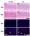
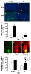
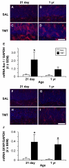
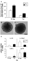
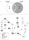
Current data suggests an association between elevations in interleukin 1 (IL-1)α, IL-1β, and IL-6 and the proliferation of neural progenitor cells (NPCs) following brain injury. A limited amount of work implicates changes in these pro-inflammatory responses with diminished NPC proliferation observed as a function of aging. In the current study, adolescent (21day-old) and 1year-old CD-1 male mice were injected with trimethyltin (TMT, 2.3mg/kg, i.p.) to produce acute apoptosis of hippocampal dentate granule cells. In this model, fewer 5-bromo-2'-deoxyuridine (BrdU)+ NPC were observed in both naive and injured adult hippocampus as compared to the corresponding number seen in adolescent mice. At 48h post-TMT, a similar level of neuronal death was observed across ages, yet activated ameboid microglia were observed in the adolescent and hypertrophic process-bearing microglia in the adult. IL-1α mRNA levels were elevated in the adolescent hippocampus; IL-6 mRNA levels were elevated in the adult. In subgranular zone (SGZ) isolated by laser-capture microdissection, IL-1β was detected but not elevated by TMT, IL-1a was elevated at both ages, while IL-6 was elevated only in the adult. Naïve NPCs isolated from the hippocampus expressed transcripts for IL-1R1, IL-6Rα, and gp130 with significantly higher levels of IL-6Rα mRNA in the adult. In vitro, IL-1α (150pg/ml) stimulated proliferation of adolescent NPCs; IL-6 (10ng/ml) inhibited proliferation of adolescent and adult NPCs. Microarray analysis of SGZ post-TMT indicated a prominence of IL-1a/IL-1R1 signaling in the adolescent and IL-6/gp130 signaling in the adult.
Published by Elsevier Inc.
Figures


Similar articles
-
Injury-induced neurogenesis: consideration of resident microglia as supportive of neural progenitor cells.Neurotox Res. 2011 Feb;19(2):341-52. doi: 10.1007/s12640-010-9199-6. Epub 2010 Jun 4. Neurotox Res. 2011. PMID: 20524106 Free PMC article.
-
The type 1 interleukin 1 receptor is not required for the death of murine hippocampal dentate granule cells and microglia activation.Brain Res. 2008 Feb 15;1194:8-20. doi: 10.1016/j.brainres.2007.11.076. Epub 2007 Dec 23. Brain Res. 2008. PMID: 18191113 Free PMC article.
-
Astrocytes support hippocampal-dependent memory and long-term potentiation via interleukin-1 signaling.Brain Behav Immun. 2011 Jul;25(5):1008-16. doi: 10.1016/j.bbi.2010.11.007. Epub 2010 Nov 17. Brain Behav Immun. 2011. PMID: 21093580
-
Age-dependent decline in neurogenesis of the hippocampus and extracellular nucleotides.Hum Cell. 2019 Apr;32(2):88-94. doi: 10.1007/s13577-019-00241-9. Epub 2019 Feb 7. Hum Cell. 2019. PMID: 30730038 Review.
-
The interaction between microglia and neural stem/precursor cells.Brain Res Bull. 2014 Oct;109:32-8. doi: 10.1016/j.brainresbull.2014.09.005. Epub 2014 Sep 22. Brain Res Bull. 2014. PMID: 25245208 Review.
Cited by
-
Role of neuroinflammation mediated potential alterations in adult neurogenesis as a factor for neuropsychiatric symptoms in Post-Acute COVID-19 syndrome-A narrative review.PeerJ. 2022 Nov 4;10:e14227. doi: 10.7717/peerj.14227. eCollection 2022. PeerJ. 2022. PMID: 36353605 Free PMC article. Review.
-
Adjudin-preconditioned neural stem cells enhance neuroprotection after ischemia reperfusion in mice.Stem Cell Res Ther. 2017 Nov 7;8(1):248. doi: 10.1186/s13287-017-0677-0. Stem Cell Res Ther. 2017. PMID: 29115993 Free PMC article.
-
Insufficient Sleep and Alzheimer's Disease: Potential Approach for Therapeutic Treatment Methods.Brain Sci. 2024 Dec 28;15(1):21. doi: 10.3390/brainsci15010021. Brain Sci. 2024. PMID: 39851389 Free PMC article. Review.
-
Interleukin-1β: a new regulator of the kynurenine pathway affecting human hippocampal neurogenesis.Neuropsychopharmacology. 2012 Mar;37(4):939-49. doi: 10.1038/npp.2011.277. Epub 2011 Nov 9. Neuropsychopharmacology. 2012. PMID: 22071871 Free PMC article.
-
Heroin-induced conditioned immunomodulation requires expression of IL-1β in the dorsal hippocampus.Brain Behav Immun. 2013 May;30:95-102. doi: 10.1016/j.bbi.2013.01.076. Epub 2013 Jan 26. Brain Behav Immun. 2013. PMID: 23357470 Free PMC article.
References
-
- Ajmone-Cat MA, Cacci E, Ragazzoni Y, Minghetti L, Biagioni S. Pro-gliogenic effect of IL-1alpha in the differentiation of embryonic neural precursor cells in vitro. J Neurochem. 2010;113:1060–1072. - PubMed
-
- Alvarez-Buylla A, Lim DA. For the long run: maintaining germinal niches in the adult brain. Neuron. 2004;41:683–686. - PubMed
-
- Banasr M, Hery M, Printemps R, Daszuta A. Serotonin-induced increases in adult cell proliferation and neurogenesis are mediated through different and common 5-HT receptor subtypes in the dentate gyrus and the subventricular zone. Neuropsychopharmacology. 2004;29:450–460. - PubMed
-
- Ben Abdallah NM, Slomianka L, Vyssotski AL, Lipp HP. Early age-related changes in adult hippocampal neurogenesis in C57 mice. Neurobiol Aging. 2010;31:151–161. - PubMed
Publication types
MeSH terms
Substances
Grants and funding
LinkOut - more resources
Full Text Sources
Other Literature Sources

