. 2010 Sep 14;19(3):377-88.
doi: 10.1016/j.devcel.2010.08.011.

Rho-kinase directs Bazooka/Par-3 planar polarity during Drosophila axis elongation

Affiliations

Rho-kinase directs Bazooka/Par-3 planar polarity during Drosophila axis elongation

Sérgio de Matos Simões et al. Dev Cell. .

Abstract

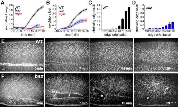
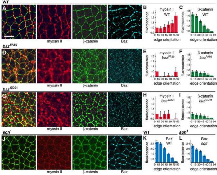
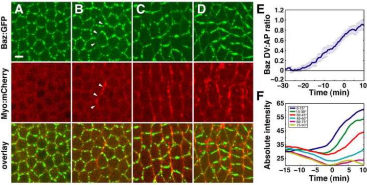
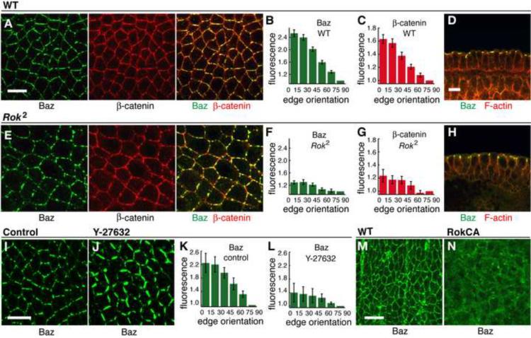
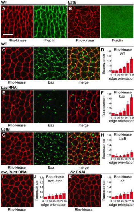
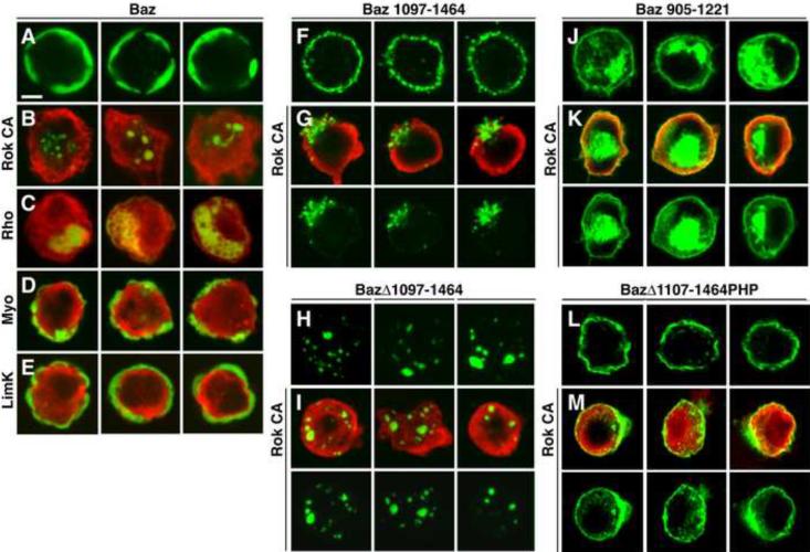
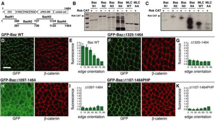
Cell rearrangements shape the Drosophila embryo via spatially regulated changes in cell shape and adhesion. We show that Bazooka/Par-3 (Baz) is required for the planar polarized distribution of myosin II and adherens junction proteins and polarized intercalary behavior is disrupted in baz mutants. The myosin II activator Rho-kinase is asymmetrically enriched at the anterior and posterior borders of intercalating cells in a pattern complementary to Baz. Loss of Rho-kinase results in expansion of the Baz domain, and activated Rho-kinase is sufficient to exclude Baz from the cortex. The planar polarized distribution of Baz requires its C-terminal domain. Rho-kinase can phosphorylate this domain and inhibit its interaction with phosphoinositide membrane lipids, suggesting a mechanism by which Rho-kinase could regulate Baz association with the cell cortex. These results demonstrate that Rho-kinase plays an instructive role in planar polarity by targeting Baz/Par-3 and myosin II to complementary cortical domains.


Figures

Figure 1
Figure 1. baz mutants have defects in cell intercalation
(A–D) Cell behavior in time-lapse movies of wild-type and mutant embryos. (A) Germband elongation is strongly reduced in bazGD21 and sqh1 mutants. (B) Fewer neighbors are lost per cell in baz and sqh mutants. An average value was obtained for each embryo, error bars indicate s.e.m. across embryos (n=10 WT, 5 bazGD21, and 2 sqh1 embryos). WT embryos were imaged with GFP:Resille (4), GFP:Spider (3), and E-cadherin:GFP (3), bazGD21 embryos were imaged with GFP:Spider, sqh1 embryos were imaged with E-cadherin:GFP. t=0 is the onset of elongation in stage 7. (C) Contractile behavior in wild type is biased toward edges perpendicular to the AP axis (angles in degrees, 0 for edges parallel to the AP axis). Bars indicate 10° bins starting at 0–10°. (D) Contractile behavior in baz mutants is reduced and partially spatially deregulated: 66% of shrinking edges were oriented at ≥60° in baz mutants compared to 88% in wild type, 16% were oriented at ≤30° in baz mutants compared to <1% in wild type (n = 10 wild-type and 5 bazGD21 embryos, 33–161 cells/embryo). (E,F) Stills from movies of wild-type (E) and bazGD21 (F) embryos expressing Spider:GFP. Asterisks indicate defects in ventral furrow closure in baz mutants and arrows point to clusters of apically constricting cells. Scale bar = 10 μm. See Figure S1, Movies S1,2.
Figure 2
Figure 2. Baz/Par-3 is required for the planar polarized localization of myosin II and β-catenin
(A,D,G,J) Localization of myosin II (Zip heavy chain, red), β-catenin (green), and Baz (blue) in stage 7 wild-type (A), bazFA50 (D), bazGD21 (G), and sqh1 (J) embryos. Left panels show overlays of myosin (red) and β-catenin (green). Anterior left, ventral down. Scale bar = 10 μm. (B,E,H) Myosin planar polarity was disrupted in bazFA50 (P<0.001) (E) and bazGD21 (P<0.001) (H) compared to wild type (B) (n=5 wild-type, 3 bazFA50, 4 bazGD21 embryos). (C,F,I) β-catenin planar polarity was disrupted in bazFA50 (P<0.001) (F) and bazGD21 (P<0.001) (I) compared to wild type (C) (n=11 wild-type, 3 bazFA50, 4 bazGD21 embryos). (K,L) Baz planar polarity was retained in sqh1 mutants (L) compared to wild type (K) (n=11 wild-type, 14 sqh1 embryos). See Figure S2.
Figure 3
Figure 3. Baz/Par-3 is downregulated at AP edges and upregulated at DV edges, establishing planar polarity
(A–D) Stills from a movie of intercalating cells in an embryo expressing Myo:mCherry (red) and Baz:GFP under the control of a Gal4 driver (green). t = 0 is the onset of elongation in stage 7. A −11 min, B −4 min, C 3 min, D 9 min. (B) In stage 6, Baz:GFP is displaced from and Myo:mCherry accumulates at AP edges (arrowheads). (C,D) In stage 7, Myo:mCherry and Baz:GFP occupy AP and DV edges, respectively. Scale bar = 5 μm. (E,F) Quantitation of Baz:GFP fluorescence intensity. (E) The log2 ratio of Baz:GFP intensity at DV edges (oriented at ≤30° relative to the AP axis) relative to AP edges (oriented at ≥60° relative to the AP axis) was averaged across all cells (n = 5 embryos, 764–2142 edges/embryo). Error bars indicate s.e.m. across embryos. t = 0 is the onset of elongation in stage 7. (F) Absolute edge intensities grouped by edge orientation in a single embryo. Baz is first downregulated at AP edges (yellow, purple) and then upregulated at DV edges (dark blue, green). See Figure S3, Movie S3.
Figure 4
Figure 4. Rho-kinase is required for Baz/Par-3 planar polarity
(A–H) Localization of Baz (green) and β-catenin (red) in stage 7 wild-type (A–D) and DRok2 mutant (E–H) embryos. Cross sections in D,H. Quantitation of Baz and Arm/β-catenin planar polarity in wild type (B,C) and DRok2 (F,G). Baz and β-catenin planar polarity were strongly reduced in DRok2 mutant embryos compared to wild type (P<0.001 for each result) (n=11 wild-type, 8 DRok2 embryos). Two classes of defects were observed, with the more defective class shown here. See Figure S4. (I–L) Localization of Baz (green) in stage 7 embryos injected with water (I) or Y-27632 (J). Baz planar polarity was reduced in Y-27632-injected embryos (K) compared to wild type (L) (P=0.002) (n=5 control injected, 4 Y-27632-injected embryos). (M,N) Localization of Baz (green) in the dorsolateral epidermis of a stage 14 control embryo (M) or an embryo expressing activated Rho-kinase (Rok-CA, N). Scale bars = 10 μm.
Figure 5
Figure 5. Rho-kinase is planar polarized in intercalating cells
(A,B) Localization of Rho-kinase (HA:Rok, red) and F-actin (phalloidin, green) in stage 7 wild-type (A) or LatB-injected (B) embryos. (C–L) Localization of Rho-kinase (Venus:RokK116A, red) and Baz (green). Stage 7 wild-type embryo (C) or embryos injected with baz dsRNA (E), LatB (G), eve and runt dsRNA (I), or Kr dsRNA (K). Quantitation of planar polarity in wild-type (D) and injected embryos (F,H,J,L). Rho-kinase planar polarity was not affected by baz RNAi (F) (n=6 wild-type Venus:RokK116A expressing embryos, 7 baz dsRNA-injected embryos, P=0.29). Rho-kinase planar polarity was reduced by injection of LatB (H) (5 embryos, P<0.001), eve and runt dsRNA (J) (7 embryos, P<0.001), or Kr dsRNA (L) (4 embryos, P=0.01). Scale bar = 10 μm.
Figure 6
Figure 6. Rho-kinase regulates the association of the Baz/Par-3 C-terminal domain with the cortex
(A–E) Drosophila S2R+ cells expressing Baz:Venus alone (green, A) or with activated versions of Rho-kinase (Rok CA red, B), Rho1 GTPase (RhoV14 red, C), myosin regulatory light chain (sqhE20,E21 red, D) or Lim kinase (limKE591 isoform C red, E). Baz:Venus was cortical in 78% of cells without Rok CA (n=216) vs. 5% with Rok CA (n=284). Expression of RhoV14, but not sqhE20,E21 or limKE591, led to a loss of Baz from the cortex. (F–K) Cells expressing Baz:Venus variants alone (green, F,H,J) or with Rok CA (red, G,I,K). (F,G) The Baz C-terminus is sufficient for cortical localization (F) and is displaced from the cortex by Rok CA (G). Baz 1097-1464 was cortical in 99% of cells without Rok CA (n=190) vs. 2% with Rok CA (n=178). (H,I) Deletion of the Baz C-terminus (BazΔ1097–1464) abolished Baz cortical localization in the absence or presence of Rok CA. (J,K) An adjacent region of the Baz C-terminus (Baz 905–1221) was not affected by Rok CA. Baz 905–1221 was cortical in 91% of cells without Rok CA (n=137) vs. 97% with Rok CA (n=208). (L,M) Cells expressing a GFP:Baz variant where the C-terminus was replaced with a heterologous PH domain. BazΔ1107–1464PHP localizes to the cortex (L) but was not efficiently displaced by Rok CA (M). GFP:Baz cortical localization was reduced 5.5-fold from 94% to 17% by Rok CA. GFP:BazΔ1107–1464PHP cortical localization was reduced 1.4-fold from 74% to 54% by Rok CA (n=183–257 cells/condition). Scale bar = 5 μm. See Figures S5,6.
Figure 7
Figure 7. The Baz/Par-3 C-terminal domain is necessary for Baz planar polarity
(A–C) GST-Baz fragments were incubated with activated Rho-kinase (Rok-CA) and γ-32P-ATP. (A) Domains in each fragment. (B) Total protein quantified by Coomassie staining. Molecular weight markers in kD (top to bottom): 100, 75, 50 and 37. (C) Phosphorylated proteins were detected by autoradiography. Arrowheads indicate Rok-CA, which shows weak autophosphorylation. Asterisks indicate GST-Baz fragments, wild-type chick myosin regulatory light chain (MRLC) (positive control), and unphosphorylatable chick MRLC T19A,S20A (negative control). (D–K) Localization of GFP-Baz transgenes in stage 7 embryos. Endogenous baz was knocked down by dsRNA injection and embryos were labeled with antibodies to GFP (green) and β-catenin (red). (D,E) Full-length GFP-Baz displays wild-type planar polarity (5 embryos). (F,G) GFP-BazΔ1325–1464 localizes to the cortex but fails to localize asymmetrically (4 embryos, P<0.001). (H,I) GFP-BazΔ1097–1464 lacking a larger region of the C-terminus shows reduced cortical association and fails to localize asymmetrically (3 embryos, P<0.001). (J,K) GFP-BazΔ1107–1464:PHP where the C-terminal 358 amino acids of Baz were replaced with a heterologous PH domain restores cortical localization but fails to localize asymmetrically (6 embryos, P<0.001). Scale bar = 10 μm. See Figure S6.

Similar articles

Cited by

References

    1. Amano M, Ito M, Kimura K, Fukata Y, Chihara K, Nakano T, Matsuura Y, Kaibuchi K. Phosphorylation and activation of myosin by Rho-associated kinase (Rho-kinase) J Biol Chem. 1996;271:20246–20249. - PubMed
    1. Amano M, Fukata Y, Kaibuchi K. Regulation and functions of Rho-associated kinase. Exp Cell Res. 2000;261:44–51. - PubMed
    1. Atwood SX, Prehoda KE. aPKC phosphorylates Miranda to polarize fate determinants during neuroblast asymmetric cell division. Curr Biol. 2009;19:723–729. - PMC - PubMed
    1. Benton R, St Johnston D. A conserved oligomerization domain in Drosophila Bazooka/PAR-3 is important for apical localization and epithelial polarity. Curr Biol. 2003;13:1330–1334. - PubMed
    1. Bertet C, Sulak L, Lecuit T. Myosin-dependent junction remodelling controls planar cell intercalation and axis elongation. Nature. 2004;429:667–671. - PubMed

Publication types

MeSH terms

LinkOut - more resources