. 2010 Sep 14;19(3):450-9.
doi: 10.1016/j.devcel.2010.08.012.

Hopx and Hdac2 interact to modulate Gata4 acetylation and embryonic cardiac myocyte proliferation

Affiliations

Hopx and Hdac2 interact to modulate Gata4 acetylation and embryonic cardiac myocyte proliferation

Chinmay M Trivedi et al. Dev Cell. .

Abstract

Regulation of chromatin structure via histone modification has recently received intense attention. Here, we demonstrate that the chromatin-modifying enzyme histone deacetylase 2 (Hdac2) functions with a small homeodomain factor, Hopx, to mediate deacetylation of Gata4, which is expressed by cardiac progenitor cells and plays critical roles in the regulation of cardiogenesis. In the absence of Hopx and Hdac2 in mouse embryos, Gata4 hyperacetylation is associated with a marked increase in cardiac myocyte proliferation, upregulation of Gata4 target genes, and perinatal lethality. Hdac2 physically interacts with Gata4, and this interaction is stabilized by Hopx. The ability of Gata4 to transactivate cell cycle genes is impaired by Hopx/Hdac2-mediated deacetylation, and this effect is abrogated by loss of Hdac2-Gata4 interaction. These results suggest that Gata4 is a nonhistone target of Hdac2-mediated deacetylation and that Hdac2, Hopx, and Gata4 coordinately regulate cardiac myocyte proliferation during embryonic development.


Figures

Figure 1
Figure 1. Myocardial defects in Hdac2-Hopx-null mice
(A) Immunohistochemistry (Hdac2) and LacZ staining (Hopx) of P0 HopxlacZ/+ heart sections shows Hdac2 and Hopx co-expression (arrow) in developing heart. (B) Wild-type and Hdac2-Hopx DKO littermates are shown at P0. (C) H&E-stained sections of wild-type and compound heterozygous hearts show muscular type of ventricular septal defect (VSD) at P0. (D) Abnormally thickened compact zone of the ventricular myocardium and increased cardiac myocyte proliferation in DKO hearts. Immunohistochemistry staining of Hdac2 shows loss of Hdac2 in DKO hearts at P0. Hopx in situ shows loss of Hopx mRNA in DKO hearts at P0. Cardiac myocyte proliferation (green, stained by MF-20) was assessed by phosphohistone H3 co-staining (arrows) in wild-type, Hdac2−/−, Hopx−/− and DKO P0 hearts. Scale bar, 20 μm. (E) Quantification of phospho-histone H3 positive cells in WT, compound heterozygous, Hdac2−/−, Hopx−/−, and DKO P0 cardiac myocytes (mean ± SEM, n=3). N.S., not significant.
Figure 2
Figure 2. Aberrant expression of Gata4 target genes in DKO hearts
(A) Transcripts for Gata4 target genes, cyclinD2 and cdk4, were detected by qRT-PCR from E16.5 wild-type, Hdac2−/−, Hopx−/− and DKO myocardium (mean ± SEM, n=3) (B) Gata4 transcripts were detected by qRT-PCR from E16.5 wild-type, Hdac2−/−, Hopx−/− and DKO myocardium (mean ± SEM, n=3). (C) Gata4 protein levels are unchanged in DKO mice. Gata4 expression was assessed by immunohistochemistry in WT and DKO E16.5 hearts. Scale bar, 10 μm. (D) Western blot analysis was performed on total lysates from WT, DKO, Hdac2−/−, and Hopx−/− E16.5 hearts. β-actin is shown as a loading control. N.S., not significant.
Figure 3
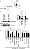
Figure 3. Hdac2-Hopx modulates Gata4 transcriptional activity and acetylation
(A) Increased Gata4 acetylation in DKO hearts. Total lysates from E16.5 WT, DKO, Hdac2−/−, and Hopx−/− hearts were immunoprecipitated by anti-Gata4 antibody and Western blot was performed using anti-acetyl lysine antibody to detect acetylated Gata4. Western blot for total Gata4 is shown below. (B) Acetylated Gata4 was quantified and normalized to total Gata4 in WT, DKO, Hdac2−/−, and Hopx−/− myocardium using ImageJ software (mean ± SEM, n=3). (C) Hdac2-Hopx over-expression inhibits Gata4 acetylation. Total lysates from transfected 293T cells were immunoprecipitated by anti-HA antibody to immunoprecipitate Gata4 and acetylated Gata4 was detected by Western blot analysis using anti-acetyl lysine antibody. (D) Acetylated Gata4 was quantified and normalized to total Gata4 using ImageJ software (mean ± SEM, n=3). (E) Hdac2-Hopx inhibits Gata4 dependent transactivation. CyclinD2-luciferase reporter construct was transfected in H9c2 myoblast cells with or without Gata4, Hdac2, Hopx, HopxH2, Gata4KA, and Gata4KQ expression constructs. Firefly luciferase activity was measured from total lysates 24 hrs after transfection and was normalized to renilla luciferase activity. The induction is represented as fold-induction over the normalized luciferase activity in the untreated cells (mean ± SEM, n=3). N.S., not significant.
Figure 4
Figure 4. Hdac2 interacts with Gata4
(A) Total lysates from E16.5 WT and Hopx−/− hearts were immunoprecipitated by anti-Gata4 antibody and Western blot was performed using anti-Hdac2 antibody. The interaction between Hdac2 and Gata4 is more robust in WT hearts (lane 2) than in Hopx-deficient hearts (lane 3). (B) Quantification of the interaction between Hdac2 and Gata4 in WT and Hopx−/− hearts using ImageJ software (mean ± SEM, n=3). (C) Hopx modulates Hdac2-Gata4 interaction. Total lysates from transfected 293T cells were immunoprecipitated by anti-HA antibody to immunoprecipitate Gata4 and Western blot was performed with anti-Flag antibody to detect Hdac2. Increasing amounts of Hopx resulted in an increasingly robust interaction between Hdac2 and Gata4. (D) Quantification of the interaction between Hdac2 and Gata4 in presence of increasing amount of Hopx (mean ± SEM, n=3). (E) Schematic representation of Gata4 showing post-translational modification sites and deletion constructs. Gata4 contains two distinct zinc (Zn) finger domains and two transcriptional activation domains (TAD). Gata4 also contains lysine rich domain (K) and nuclear localization sequence (NLS). p300 dependent acetylation of Gata4 at Lys 311, 318, 320, and 322 and MAPK dependent phosphorylation at Ser105 is important for Gata4 DNA binding and transcriptional activation abilities. (F) Total lysates from Gata4 or various Gata4 deletion constructs transfected 293T cells were immunoprecipitated with anti-HA antibody and Westernblot analysis was performed with anti-Hdac2 antibody to detect Hdac2. n.s. – non specific. (G) Loss of Hdac2 and Gata4 interaction prevents Hdac2-Hopx mediated repression of Gata4 dependent cyclinD2 reporter activity. CyclinD2-luciferase reporter construct was transfected in H9c2 cells with or without Gata4, Gata4 Δ241-274, Hdac2, and Hopx expression constructs. Firefly luciferase activity was measured from total lysates 24 hrs after transfection and was normalized to renilla luciferase activity. The induction is represented as % activity of cyclinD2 reporter over the normalized luciferase activity in the Gata4 or Gata4 Δ241-274 transfected cells (mean ± SEM, n=3).
Figure 5
Figure 5. Phosphorylated Hdac2 preferentially interacts with Gata4 and modulates transcriptional activity
(A) Total lysates from transfected 293T cells were immunoprecipitated with anti-Hdac2 antibody to immunoprecipitate Hdac2 and Western blot analysis was performed with anti-Gata4 antibody. Mutation of serine 394, 411, 422 and 424 of Hdac2 to alanine (Hdac2SA) reduced interaction with Gata4. (B) Quantification of the interaction between phospho-mutant Hdac2 and Gata4 using ImageJ software (mean ± SEM, n=3). (C) CyclinD2-luciferase reporter construct was transfected in H9c2 myoblast cells with or without Gata4, Hdac2, Hopx, and Hdac2SA expression constructs. Normalized luciferase activity is reported as fold-induction over mock transfected cells (mean ± SEM, n=3). (D) Hdac2SA retains deacetylase activity. Total lysates from transfected 293T cells with or without Hdac2 and Hdac2SA were assayed for HDAC activity against a pseudo-substrate (mean ± SEM, n=3). (E) Western blot analysis was performed on total lystates from transfected 293T cells with or without Hdac2 or Hdac2SA using an anti-acetylated histoneH3 antibody. Hdac2 expression was determined by using an anti-Hdac2 antibody that recognized endogenous (lane 1) and exogenous Hdac2. Transfection of either Hdac2 or Hdac2SA resulted in diminished histoneH3 acetylation compared to mock transfected cells. (F) Acetylated histoneH3 was quantified and normalized to total histoneH3 in Hdac2 or Hdac2SA transfected cells using ImageJ software (mean ± SEM, n=3). (G) Model depicting Hdac2 interacting with Hopx to induce deacetylation of Gata4 and modulation of cell cycle genes.

References

    1. Adenuga D, Yao H, March TH, Seagrave J, Rahman I. Histone deacetylase 2 is phosphorylated, ubiquitinated, and degraded by cigarette smoke. Am J Respir Cell Mol Biol. 2009;40:464–473. - PMC - PubMed
    1. Arceci RJ, King AA, Simon MC, Orkin SH, Wilson DB. Mouse GATA-4: a retinoic acid-inducible GATA-binding transcription factor expressed in endodermally derived tissues and heart. Mol Cell Biol. 1993;13:2235–2246. - PMC - PubMed
    1. Barrett T, Troup DB, Wilhite SE, Ledoux P, Rudnev D, Evangelista C, Kim IF, Soboleva A, Tomashevsky M, Marshall KA, et al. NCBI GEO: archive for high-throughput functional genomic data. Nucleic Acids Res. 2009;37:D885–890. - PMC - PubMed
    1. Cantor AB, Orkin SH. Coregulation of GATA factors by the Friend of GATA (FOG) family of multitype zinc finger proteins. Semin Cell Dev Biol. 2005;16:117–128. - PubMed
    1. Charron F, Nemer M. GATA transcription factors and cardiac development. Semin Cell Dev Biol. 1999;10:85–91. - PubMed

Publication types

MeSH terms

Associated data