. 2010 Sep 16;8(3):225-35.
doi: 10.1016/j.chom.2010.08.002.

A biphasic innate immune MAPK response discriminates between the yeast and hyphal forms of Candida albicans in epithelial cells

Affiliations

A biphasic innate immune MAPK response discriminates between the yeast and hyphal forms of Candida albicans in epithelial cells

David L Moyes et al. Cell Host Microbe. .

Abstract

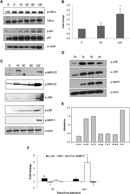
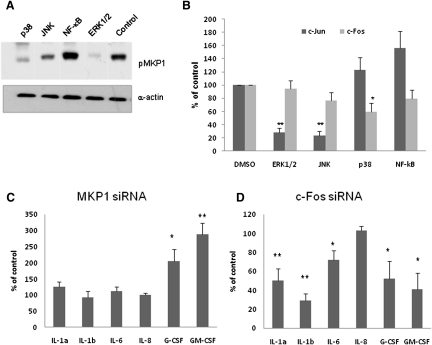
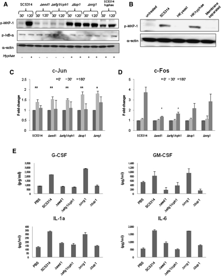
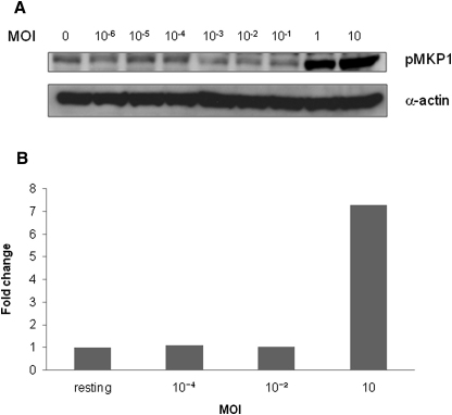
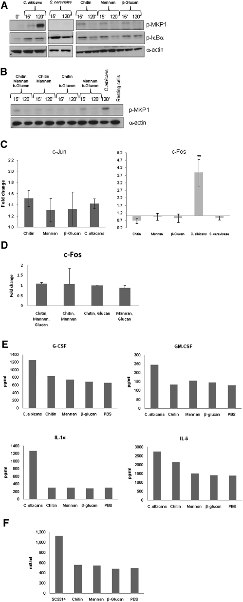
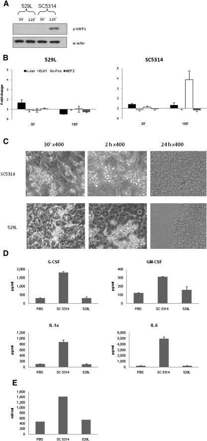
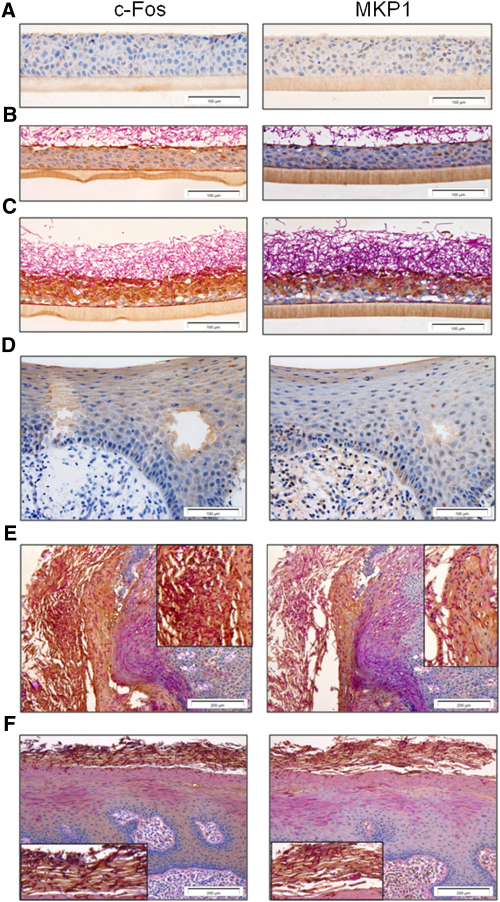
Discriminating between commensal and pathogenic states of opportunistic pathogens is critical for host mucosal defense and homeostasis. The opportunistic human fungal pathogen Candida albicans is also a constituent of the normal oral flora and grows either as yeasts or hyphae. We demonstrate that oral epithelial cells orchestrate an innate response to C. albicans via NF-κB and a biphasic MAPK response. Activation of NF-κB and the first MAPK phase, constituting c-Jun activation, is independent of morphology and due to fungal cell wall recognition. Activation of the second MAPK phase, constituting MKP1 and c-Fos activation, is dependent upon hypha formation and fungal burdens and correlates with proinflammatory responses. Such biphasic response may allow epithelial tissues to remain quiescent under low fungal burdens while responding specifically and strongly to damage-inducing hyphae when burdens increase. MAPK/MKP1/c-Fos activation may represent a "danger response" pathway that is critical for identifying and responding to the pathogenic switch of commensal microbes.


Figures

Figure 1
Figure 1
C. albicans Infection of TR146 Epithelial Cells Activates NF-κB and MAPK Signaling (A) Increasing phosphorylation of IκB-α and p65 from 5 min postinfection onward. Bands are shown relative to unphosphorylated protein and α-actin loading control. (B) Transcription factor DNA binding activity of p65 after C. albicans infection as measured by TransAM ELISA. (C) Increased phosphorylation of MEK1/2, ERK1/2, JNK, and p38 after 15 min postinfection, with further phosphorylation at 2 hr, indicating a biphasic response. Also, phosphorylation of MKP1 only at 2 hr postinfection, indicating stabilization of the MAPK response. (D) Decreasing levels of p38 and JNK phosphorylation after 2 hr infection, matching with increasing levels of MKP1 phosphorylation. (E) Levels of DNA binding activity (absorbance values) of AP-1 transcription factor members in resting TR146 cells. (F) Changes in binding activity of c-Fos, c-Jun, Elk1, and MEF2 after infection; data represented as fold change relative to resting cells. A C. albicans:epithelial cell MOI of 10:1 was used. Data are (A, C–E) representative of three independent experiments or mean of at least three (B) or six (F) independent experiments ± SEM. p < 0.05, ∗∗p < 0.01. (Two bands are seen for ERK1/2 and JNK, as two different proteins make up these complexes). See also Figure S1.
Figure 2
Figure 2
Regulation of MKP1, c-jun, c-Fos, and Cytokine Activation (A) Levels of MKP1 phosphorylation 2 hr after C. albicans infection (MOI = 10) in TR146 cells after inhibition of different pathways. (B) Level of inhibition of c-Jun (30 min) and c-Fos (3 hr) DNA binding activity after pretreatment for 4 hr with signaling pathway inhibitors. (C and D) Effect of MKP1 and c-Fos knockdown, respectively, on cytokine production (MOI of 0.01). Data are expressed as percentage of vehicle control levels (B–D) and are representative of three independent experiments (A) or are the mean of four independent experiments ± SEM (B–D). p < 0.05; ∗∗p < 0.01; ∗∗∗p < 0.001. See also Figure S2.
Figure 3
Figure 3
Specific Activation of MKP1 and Transcription Factors and Cytokine Induction by C. albicans (A) Immunoblot of phosphorylated MKP1 and IκB-α after infection of TR146 cells with wild-type (SC5314), hyperfilamentous (Δtup1 and Δnrg1), and nonfilamentous (Δeed1 and Δefg1/cph1) strains or pregrown C. albicans hyphae. The morphological status of the strains at each time point is indicated below the immunoblots (presence [+] or absence [−] of filaments). (B–E) (B) Induction of MKP1 phosphorylation at 2 hr using heat-killed (HK) wild-type (SC5314) yeast or hyphae or by SC5314 prevented from direct contact with epithelial cells by a 0.4 μm membrane. We also show changes in binding activity of c-Jun (C) and c-Fos (D) at 30 min and 3 hr infection with nonfilamentous (Δeed1 and Δefg1/cph1) or hyperfilamentous (Δtup1 and Δnrg1) mutants. (E) Induction of cytokines after 24 hr infection with nonfilamentous (Δeed1 and Δefg1/cph1) or hyperfilamentous (Δtup1 and Δnrg1) mutants. GM-CSF production appears to decrease, but this was not significant or reproduced with the other cytokines. An MOI of 10 was used for infections in (A–D), and an MOI of 0.01 was used for (E). Data are (A and B) representative of three independent experiments, (C and D) mean of four experiments (± SEM) or (E) representative of three experiments. p < 0.05; ∗∗p < 0.01. See also Figure S3.
Figure 4
Figure 4
Effect of Fungal Burdens on MKP1 and c-Fos Activation (A and B) Induction of (A) MKP1 phosphorylation and (B) c-Fos DNA binding activity by differing fungal burdens. An MOI of 1 or higher is required to activate the second MAPK phase. Wild-type SC5314 C. albicans was added as yeast cells at each MOI, and infections were left for (A) 2 hr or (B) 3 hr in each case. Data are representative of three independent experiments.
Figure 5
Figure 5
Activation of MKP1 and c-Fos by C. albicans and S. cerevisiae and Epithelial Responses to Fungal Cell Wall Components (A–D) (A and B) Phosphorylation of MKP1 and IκBα after 2 hr or (C and D) activation of c-Jun and c-Fos DNA binding activity after 30 min and 3 hr of infection, respectively, with either C. albicans or S. cerevisiae or treatment with 2 μg/ml C. albicans chitin, 50 μg/ml C. albicans N- and O- mannan, or 100 μg/ml β-glucan microspheres (individually or in combination) for the same time periods. Only C. albicans activates MKP1 and c-Fos. (C and D) Data are ± SEM. (E) Moiety-induced cytokine production after 24 hr as measured by multiplex microbead assay. (F) Moiety-induced damage as measured by LDH release after 24 hr. Doses for (B)–(D) were the same as those for (A). All experiments are (A and B) representative of or (C–E) the mean of at least three independent experiments. An MOI of 10 was used for both species. p < 0.05; ∗∗p < 0.01. See also Figure S4.
Figure 6
Figure 6
Activation of the Biphasic MAPK Response May Be Required for Colonization in a Murine Model (A and B) (A) Immunoblot analysis of MKP1 phosphorylation and (B) changes in transcription factor binding activity after infection with C. albicans SC5314 (noncolonizing) and 529L (colonizing). (C) Morphology of C. albicans strains on epithelial cells after 30 min, 2 hr, and 24 hr. (D) Secretion of cytokines after infection with C. albicans 529L and SC5314 for 24 hr. An MOI of 10 was used for (A) and (B) and an MOI of 0.01 for (D) and (E). Data are (A and C) representative of three independent experiments or (B, D, and E) mean of three independent experiments ± SEM.
Figure 7
Figure 7
Expression of c-Fos and MKP1 in Oral Epithelium (A–C) (A) Resting expression of c-Fos and MKP1 in oral RHE. Upregulation of c-Fos and MKP1 expression is associated with contact with Candida hyphae at (B) the surface after 4 hr and (C) throughout the epithelial layer when hyphae penetrate and invade at 24 hr (dark-brown staining). (D) Resting expression levels of c-Fos and MKP1 in control biopsies of human oral epithelium. (E and F) Increased expression of both c-Fos and MKP1 in two different oral biopsies with Candida infection (E, left, and F, top; dark-brown staining). Insets in (E) and (F) show an enlarged view of the region of Candida infection in each respective biopsy.

Comment in

References

    1. Abreu M.T., Fukata M., Arditi M. TLR signaling in the gut in health and disease. J. Immunol. 2005;174:4453–4460. - PubMed
    1. Agrawal S., Agrawal A., Doughty B., Gerwitz A., Blenis J., Van Dyke T., Pulendran B. Cutting edge: different Toll-like receptor agonists instruct dendritic cells to induce distinct Th responses via differential modulation of extracellular signal-regulated kinase-mitogen-activated protein kinase and c-Fos. J. Immunol. 2003;171:4984–4989. - PubMed
    1. Braun B.R., Johnson A.D. Control of filament formation in Candida albicans by the transcriptional repressor TUP1. Science. 1997;277:105–109. - PubMed
    1. Brondello J.M., Pouysségur J., McKenzie F.R. Reduced MAP kinase phosphatase-1 degradation after p42/p44MAPK-dependent phosphorylation. Science. 1999;286:2514–2517. - PubMed
    1. Chambellan A., Leahy R., Xu W., Cruickshank P.J., Janocha A., Szabo K., Cannady S.B., Comhair S.A., Erzurum S.C. Pivotal role of c-Fos in nitric oxide synthase 2 expression in airway epithelial cells. Nitric Oxide. 2009;20:143–149. - PMC - PubMed

Publication types

MeSH terms