. 2010 Sep 16;8(3):260-70.
doi: 10.1016/j.chom.2010.08.003.

In vivo mechanisms of vaccine-induced protection against HPV infection

Affiliations

In vivo mechanisms of vaccine-induced protection against HPV infection

Patricia M Day et al. Cell Host Microbe. .

Abstract

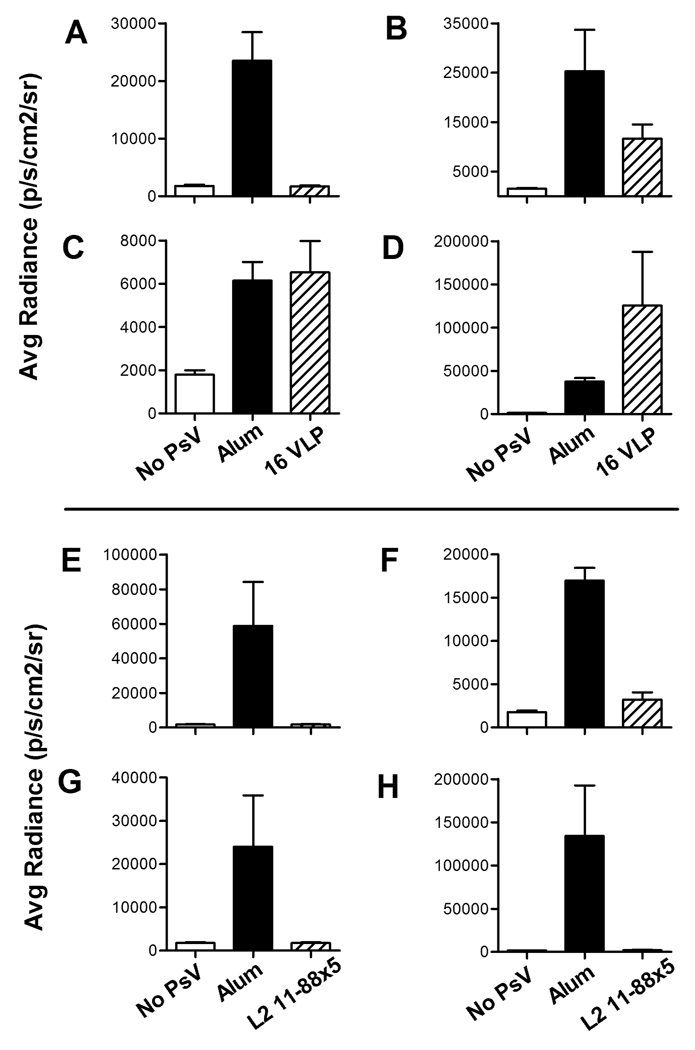
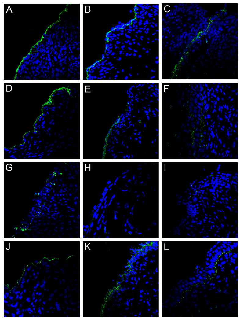
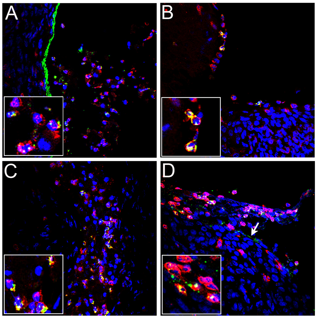
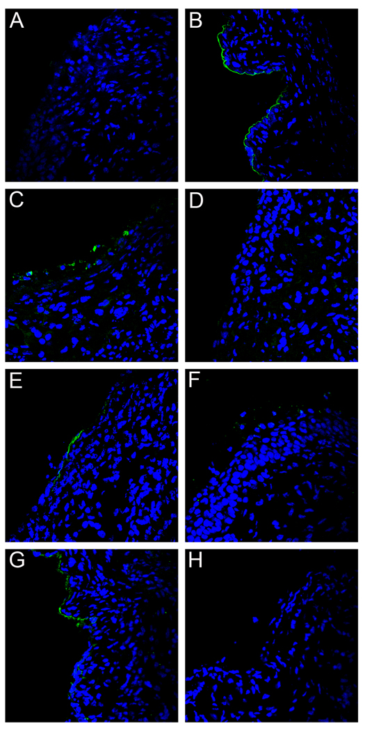
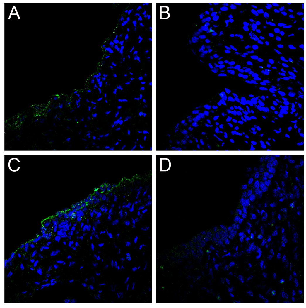
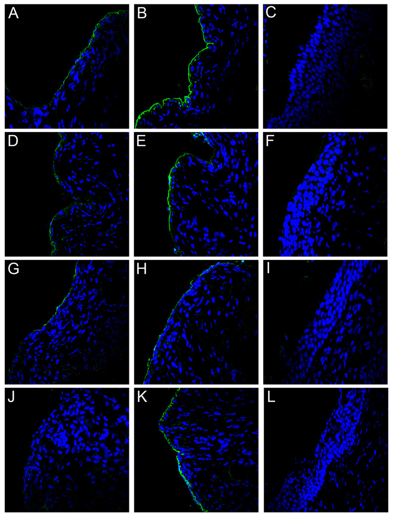
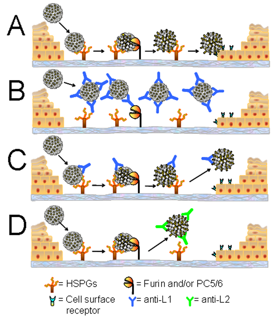
Using a human papillomavirus (HPV) cervicovaginal murine challenge model, we microscopically examined the in vivo mechanisms of L1 virus-like particle (VLP) and L2 vaccine-induced inhibition of infection. In vivo HPV infection requires an initial association with the acellular basement membrane (BM) to induce conformational changes in the virion that permit its association with the keratinocyte cell surface. By passive transfer of immune serum, we determined that anti-L1 antibodies can interfere with infection at two stages. Similarly to active VLP immunization, transfer of high L1 antibody concentrations prevented BM binding. However, in the presence of low concentrations of anti-L1, virions associated with the BM, but to the epithelial cell surface was not detected. Regardless of the concentration, L2 vaccine-induced antibodies allow BM association but prevent association with the cell surface. Thus, we have revealed distinct mechanisms of vaccine-induced inhibition of virus infection in vivo.


Figures

Figure 1
Figure 1. Protection from infection after HPV16 L1 VLP or L2 11–88x5 vaccination
Mice were vaccinated with Alum only (solid bars), 5µg HPV16 L1 VLPs or 25µg L2 11–88x5 (hatched bars) and challenged with (A, E) HPV16, (B, F) HPV31, (C, G) HPV45 or (D, H) HPV58. Unvaccinated mice were challenged with vehicle only (CMC; open bars) to establish background luminescence. Values on y-axis are average radiance (p/s/cm2/sr). Data represent the mean +/− SEM of five mice/group from a representative experiment.
Figure 2
Figure 2. PsV capsid localization and L2 exposure following VLP immunization
Pseudovirion capsids were evaluated for their ability to interact with the BM and epithelium and expose the L2 cross-neutralization epitope following generation of an antibody response via immunization with either adjuvant only or HPV16 VLPs and adjuvant. HPV16 pseudovirions in tissue from alum-immunized animals are shown in panels A–C. Panels A and C show the localization of AF488-coupled capsids (green) at 4 hours and 18 hours, respectively. Panel B shows the exposure of the L2 17–36 epitope at 4 hours following delivery of uncoupled pseudovirions, detected with a rabbit polyclonal antiserum against L2 a.a. 17/36 (green). Detection of HPV45 capsids is shown in panels D–F. AF488-coupled capsids are shown in panels D and F at 4 hours and 18 hours, respectively. The L2 epitope exposure at 4 hours is shown in panel E. The remaining panels show the analogous experiments performed in the VLP-immunized animals. HPV16 pseudovirion detection is shown in panels G–I. Capsid binding is shown for the two time points in panels G and I. L2 epitope exposure at 4 hours is shown in panel H. HPV45 pseudovirus binding is shown in panels J–L. Capsid association is in panels J and L at 4 hours and 18 hours. L2 exposure is in panel K. Images are representative of five animals tested for each condition examined.
Figure 3
Figure 3. Colocalization of pseudovirus with neutrophils
HPV16 pseudovirus was instilled into the vaginal tract of an alum-vaccinated mouse (panel A), an HPV16 VLP-vaccinated mouse (panel B), or a mice that received passively-transferred rabbit anti-L1 immune serum (panels C, high volume and D, low volume). Alexa Fluor 488-coupled pseudovirus (green) was used in the animals shown in panels A and B. In panels C and D, the antibody-bound pseudovirus was detected with Alexa Fluor 488-coupled donkey anti-rabbit secondary antibody (green). BM-associated virions evident in panel D are indicated with the arrow. Neutrophils were detected in all panels with a rat anti-neutrophil antibody and Alexa Fluor 594-coupled donkey anti-rat secondary antibody (red). All tissues were harvested at 4 hours post-instillation. Images are representative of five animals tested for each condition examined.
Figure 4
Figure 4. Localization of capsids following passive transfer of anti-VLP sera
Either pre-immune or rabbit anti-HPV16 VLP immune sera was passively transferred into mice and the localization of capsids analyzed at either 4 or 18 hours following delivery. The capsid-antibody complexes were detected with Alexa Fluor 488-conjugated donkey anti-rabbit (green). Detection of binding of pre-immune sera at 4 hours is shown in panel A. Detection of capsids in the same tissue with an anti-HPV16 L1 serum is shown in panel B. Detection of capsid-antibody complexes following the transfer of a high volume of immune serum is shown in panels C (4 hrs.) and D (18 hrs.). Detection of capsid-antibody complexes following the transfer of a low volume of immune serum is shown in panels E (4 hrs.) and F (18 hrs.). Detection of exposure of L2 following low volume passive transfer is shown in panel G (4 hrs.). This detection required blocking the passively transferred rabbit serum with unlabeled donkey anti-rabbit serum prior to binding of the rabbit anti-L2 serum which was detected with an Alexa Fluor 488-conjugated donkey anti-rabbit secondary antibody (green). Complete blocking of the low volume passive transfer was achieved. The omission of the anti-L2 serum, but inclusion of the Alexa Fluor 488-conjugated donkey anti-rabbit secondary antibody, resulted in no detectable signal (panel H). The experiment could not be performed with the high volume passive transfer due to the inability to achieve complete blocking (data not shown). Images are representative of five animals tested for each condition examined. See also Figures S1, S2, S3.
Figure 5
Figure 5. Capsid localization and L2 exposure following L2 immunization
Capsid binding and localization was examined in mice that had been immunized with the L2 11–88x5. HPV16 capsid binding was examined at 4 hours (panel A) and 18 hours (panel B) post-instillation (green). Likewise, HPV45 capsid binding was examined at 4 hours (panel C) and 18 hours (panel D). Rabbit anti-L1 serum against the appropriate type was used for staining in order to show capsid localization. Images are representative of five animals tested for each condition examined.
Figure 6
Figure 6. Localization of capsids following passive transfer of anti-L2 sera
Rabbit anti-L2 immune serum was passively transferred into mice and the localization of capsids analyzed at either 4 hours (panels A, B, D, E, G, H, and K) or 18 hours (panels C, F, I, and L) following delivery. Panels A–F show localization of HPV16. Panels G–L show localization of HPV45. The capsid-antibody complexes at 4 hours were detected with Alexa Fluor 488-conjugated donkey anti-rabbit (green) in panels A, D, G and J, showing weak staining of the antibody-capsid complexes following either transfer of high volume (panels A and G) or low volume (panel D) of immune serum. Visualization of anti-L2 bound to HPV45 capsids in the low volume transfer was below the threshold of detection (panel J). Detection of capsids (instead of antibody-bound capsids) with anti-L1 serum is shown for the same sections in the adjacent panels (B, E, H and K). Detection of capsids with the anti-L1 serum at 18 hours is shown in panel C for high volume, HPV16, panel F for low volume, HPV16, panel I for high volume, HPV45, and panel L for low volume, HPV45. Images are representative of five animals tested for each condition examined. See also Figure S1.
Figure 7
Figure 7. Model of HPV binding and interference by vaccine-induced antibodies
Panel A depicts normal in vivo binding, cleavage, and transfer of the virion to the epithelial cell surface. BM binding is blocked in the presence of high concentrations of anti-L1 antibodies (B), however at low concentrations (C), anti-L1 antibodies prevent stable engagement of the cell surface receptor. Anti-L2 antibodies bind only after the initial conformation change on the BM and cleavage of L2 by furin or PC5/6 (D) and also prevent stable binding to the cell surface.

Comment in

  • Stopping HPVs dead in their tracts.
    Carter JJ, Galloway DA. Carter JJ, et al. Cell Host Microbe. 2010 Sep 16;8(3):221-2. doi: 10.1016/j.chom.2010.08.010. Cell Host Microbe. 2010. PMID: 20833372

Similar articles

Cited by

References

    1. Bernard H-U, Burk RD, Chen Z, van Doorslaer K, Hausen Hz, de Villiers E-M. Classification of papillomaviruses (PVs) based on 189 PV types and proposal of taxonomic amendments. Virology. 2010;401:70–79. - PMC - PubMed
    1. Breitburd F, Kirnbauer R, Hubbert N, Nonnenmacher B, Trin-Dinh-Desmarquet C, Orth G, Schiller J, Lowy D. Immunization with viruslike particles from cottontail rabbit papillomavirus (CRPV) can protect against experimental CRPV infection. J. Virol. 1995;69:3959–3963. - PMC - PubMed
    1. Brown DR, Kjaer SK, Sigurdsson K, Iversen OE, Hernandez-Avila M, Wheeler CM, Perez G, Koutsky LA, Tay EH, Garcia P, et al. The Impact of Quadrivalent Human Papillomavirus (HPV; Types 6, 11, 16, and 18) L1 Virus-Like Particle Vaccine on Infection and Disease Due to Oncogenic Nonvaccine HPV Types in Generally HPV-Naive Women Aged 16–26 Years. The Journal of Infectious Diseases. 2009;199:926–935. - PubMed
    1. Buck CB, Thompson CD. Production of papillomavirus-based gene transfer vectors. Curr Protoc Cell Biol. 2007;Chapter 26(Unit 26 21) - PubMed
    1. Carter JJ, Wipf GC, Madeleine MM, Schwartz SM, Koutsky LA, Galloway DA. Identification of Human Papillomavirus Type 16 L1 Surface Loops Required for Neutralization by Human Sera. J. Virol. 2006;80:4664–4672. - PMC - PubMed

Publication types

MeSH terms

Substances