. 2010 Sep 16;8(3):271-83.
doi: 10.1016/j.chom.2010.08.007.

The human immune response to Dengue virus is dominated by highly cross-reactive antibodies endowed with neutralizing and enhancing activity

Affiliations

The human immune response to Dengue virus is dominated by highly cross-reactive antibodies endowed with neutralizing and enhancing activity

Martina Beltramello et al. Cell Host Microbe. .

Abstract

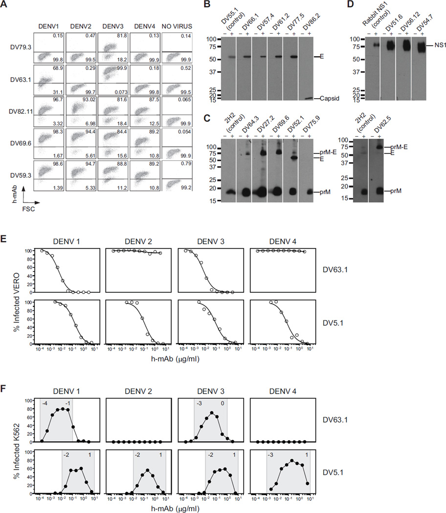
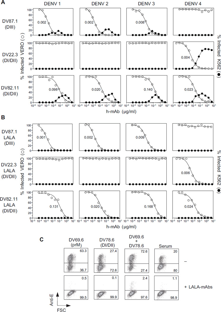
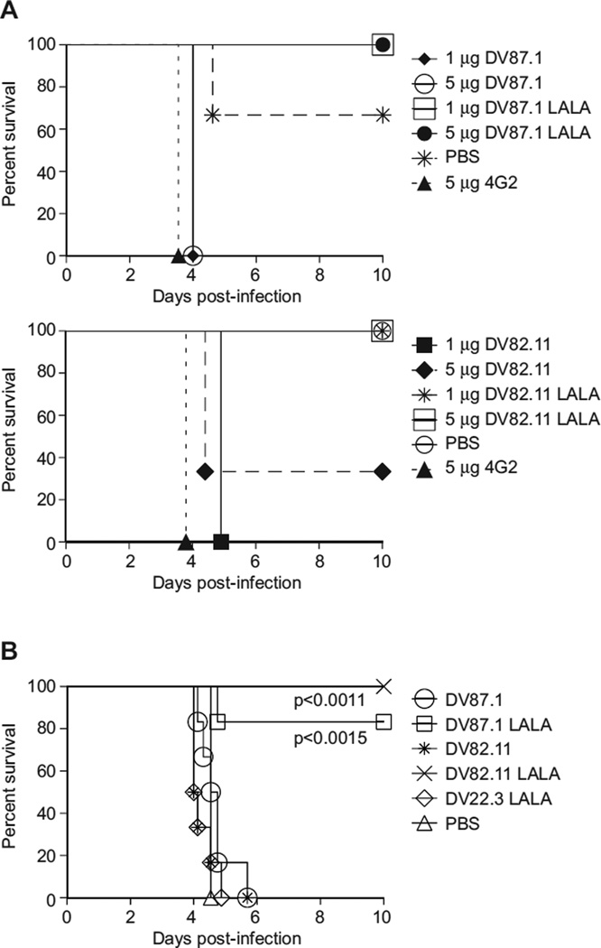
Antibodies protect against homologous Dengue virus (DENV) infection but can precipitate severe dengue by promoting heterotypic virus entry via Fcγ receptors (FcγR). We immortalized memory B cells from individuals after primary or secondary infection and analyzed anti-DENV monoclonal antibodies (mAbs) thus generated. MAbs to envelope (E) protein domain III (DIII) were either serotype specific or cross-reactive and potently neutralized DENV infection. DI/DII- or viral membrane protein prM-reactive mAbs neutralized poorly and showed broad cross-reactivity with the four DENV serotypes. All mAbs enhanced infection at subneutralizing concentrations. Three mAbs targeting distinct epitopes on the four DENV serotypes and engineered to prevent FcγR binding did not enhance infection and neutralized DENV in vitro and in vivo as postexposure therapy in a mouse model of lethal DENV infection. Our findings reveal an unexpected degree of cross-reactivity in human antibodies against DENV and illustrate the potential for an antibody-based therapy to control severe dengue.


Conflict of interest statement

The other authors have no conflicting financial interests.

Figures

Figure 1
Figure 1. Specificity and function of human mAbs against DENV
(A) Vero cells, uninfected or infected by DENV-1, −2, −3, and −4 vaccine strains, were fixed, permeabilized, and stained with human mAbs, followed by fluorescently-labeled anti-human IgG polyclonal antibodies. Representative dot plots show staining by mAbs isolated from Donor 76 reactive against DENV-3 alone (DV79.3), DENV-1 and DENV-3 (DV63.1) or cross-reactive against the four DENV serotypes (DV82.11, DV69.6, DV59.3). Numbers in quadrants indicate the percentage of positive cells. The data shown are for five mAbs and are representative of data obtained with 70 DENV-reactive mAbs and of at least two independent experiments. (B–D) Western blot analysis of mAbs that were not reactive by ELISA. Concentrated supernatants of uninfected control (−) or DENV-3-infected Vero cells (+) were separated under non-reducing conditions. B) E- and capsid-specific mAbs. E protein was identified by DV55.1, a mAb specific for E protein in ELISA. Capsid migrate with apparent molecular weights of 16 kD. C) shows prM-specific mAbs. PrM protein was identified by the mouse anti-prM mAb 2H2. D) shows NS1-specific mAbs. NS1 protein was identified by a rabbit anti-NS1 serum. The data are representative of at least two independent experiments. (E) Examples of viral neutralization assay in which serial dilutions of mAbs (DV63.1 and DV5.1) were incubated with DENV-1-4 before addition to Vero cells. Shown is the percentage of infected cells after 3 days as a function of increasing mAb concentration. Nonlinear regression analysis was used to calculate the EC50, values indicated in the panel. (F) Infection enhancement assay in which serial mAb dilutions were incubated with DENV-1-4 before addition to K562 cells. Shown is the percentage of infected K562 cells. The range of mAb concentrations where infection enhancement was observed is indicated in gray. Numbers in the gray areas represent logarithm power. The data shown are from one representative experiment out of three independent experiments performed. Data obtained with the panel of DENV-reactive mAbs are shown in Tables 2, 3 and 4.
Figure 2
Figure 2. Three mAbs neutralize all four DENV serotypes by binding two distinct epitopes on each virus
Three mAbs (all IgG1) were selected for their specificity and neutralizing activity: DV87.1 binds DIII of DENV-1, −2 and −3; DV22.3 binds DI/DII of DENV-4, and DV82.11 cross-reacts with DI/DII of all DENV serotypes. (A) Neutralization (○) and enhancement (●) of infection by recombinant unmodified mAbs. (B) Neutralization (○) and enhancement (●) of infection by engineered LALA variant mAbs. Nonlinear regression analysis was used to calculate the EC50, indicated in the panel. Data are mean of duplicates. One representative experiment out of three performed. (C) A cocktail of LALA-mAbs (DV82.11= 2 µg/ml, DV87.1=DV22.3= 0.2 µg/ml) was used in combination with mAbs directed against prM (DV69.6) or DI/DII E protein (DV78.6) or serum from a primary DENV-infected donor at potent enhancing concentration (0.1 µg/ml) on K562 cells. Numbers in quadrants indicate the percentage of infected cells, as revealed by staining with an anti-E mouse mAb. The data shown are from one experiment representative of two performed. See also Figure S1 and Table S2.
Figure 3
Figure 3. LALA variants do not enhance DENV in vivo and demonstrate post-exposure therapeutic efficacy
A (Top). 1 or 5 µg of DV87.1, DV87.1 LALA variant, 4G2 or PBS were transferred i.p. in 200 µl volume into AG129 mice (n = 3 per group). The mice were infected 18–24 h later with 106 pfu DENV-2 strain D2S10. (Bottom). 1 or 5 mg of DV82.11, DV82.11 LALA variant, 4G2 or PBS were transferred i.p. in 200 µl volume into AG129 mice (n=3 per group). The mice were subsequently infected 18 to 24 h later with 106 pfu DENV-2 D2S10. All mice receiving LALA-variant mAbs survived as compared to mice receiving a lethal, antibody-enhanced infection (1 or 5 µg DV87.1 LALA, p< 0.0253; 1 or 5 µg DV82.11 LALA, p< 0.0455 compared to 4G2 mAb positive control). (B) AG129 mice were administered 35 µl anti-DENV-1 serum i.p. and were infected 24 h later with 105 pfu of DENV-2 D2S10 i.v.. Twenty-four h after infection, the mice were treated with 50 µg of either DV87.1, n=6; DV87.1 LALA, n=6; DV82.11, n=6; DV82.11 LALA, n=5; DV22.3 LALA, n=6; or PBS, n=6 i.p.. In all cases, mortality was monitored daily for ten days. Mice treated with either 50 µg DV87.1 LALA (p<0.0015) or DV82.11 LALA (p<0.011) 24 h post-infection survived significantly longer than mice receiving 50 µg of non-binding LALA variant mAb DV22.3. See also Figure S2.

References

    1. Balsitis SJ, Williams KL, Lachica R, Flores D, Kyle JL, Mehlhop E, Johnson S, Diamond MS, Beatty PR, Harris E. Lethal antibody enhancement of dengue disease in mice is prevented by fc modification. PLoS Pathog. 2010;6:e1000790. - PMC - PubMed
    1. Beasley DW, Barrett AD. Identification of neutralizing epitopes within structural domain III of the West Nile virus envelope protein. J Virol. 2002;76:13097–13100. - PMC - PubMed
    1. Bhardwaj S, Holbrook M, Shope RE, Barrett AD, Watowich SJ. Biophysical characterization and vector-specific antagonist activity of domain III of the tick-borne flavivirus envelope protein. J Virol. 2001;75:4002–4007. - PMC - PubMed
    1. Chau TN, Hieu NT, Anders KL, Wolbers M, Lien le B, Hieu LT, Hien TT, Hung NT, Farrar J, Whitehead S, Simmons CP. Dengue virus infections and maternal antibody decay in a prospective birth cohort study of Vietnamese infants. J Infect Dis. 2009;200:1893–1900. - PMC - PubMed
    1. Chau TN, Quyen NT, Thuy TT, Tuan NM, Hoang DM, Dung NT, Lien le B, Quy NT, Hieu NT, Hieu LT, et al. Dengue in Vietnamese infants--results of infection-enhancement assays correlate with age-related disease epidemiology, and cellular immune responses correlate with disease severity. J Infect Dis. 2008;198:516–524. - PMC - PubMed

Publication types