

Cerebellar hemorrhage on magnetic resonance imaging in preterm newborns associated with abnormal neurologic outcome
- PMID: 20833401
- PMCID: PMC3010295
- DOI: 10.1016/j.jpeds.2010.07.049
Cerebellar hemorrhage on magnetic resonance imaging in preterm newborns associated with abnormal neurologic outcome
Abstract
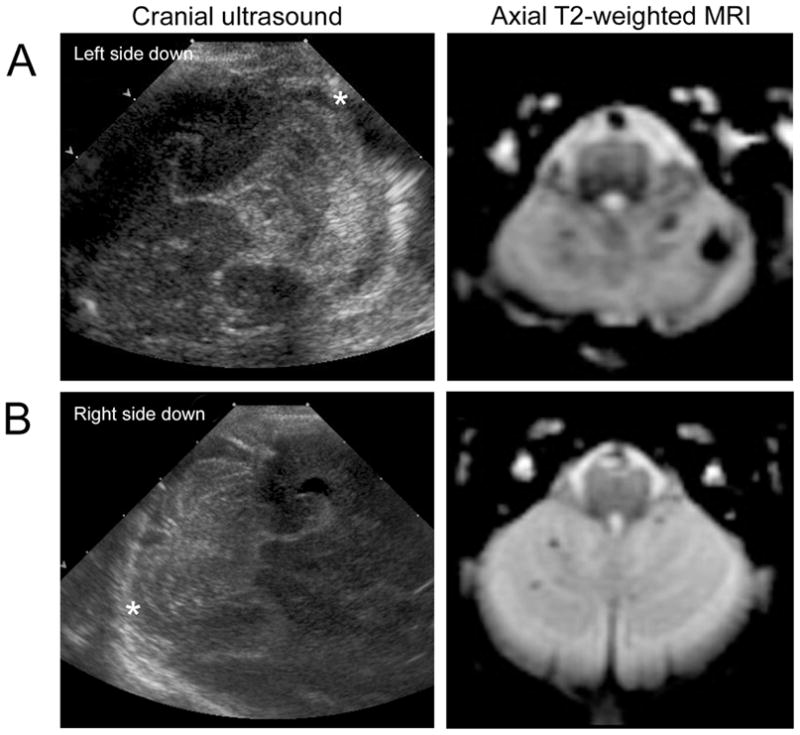
Objective: To investigate the relationship between cerebellar hemorrhage in preterm infants seen on magnetic resonance imaging (MRI), but not on ultrasonography, and neurodevelopmental outcome.
Study design: Images from a cohort study of MRI in preterm newborns were reviewed for cerebellar hemorrhage. The children were assessed at a mean age of 4.8 years with neurologic examination and developmental testing using the Wechsler Preschool and Primary Scale of Intelligence, Third Edition.
Results: Cerebellar hemorrhage was detected on both ultrasonography and MRI in 3 of the 131 preterm newborns evaluated, whereas smaller hemorrhages were seen only on MRI in 10 newborns (total incidence, 10%). Adjusting for gestational age at birth, intraventricular hemorrhage, and white matter injury, cerebellar hemorrhage detectable solely by MRI was associated with a 5-fold increased odds of abnormal neurologic examination compared with newborns without cerebellar hemorrhage (outcome data in 74%). No association with the Wechsler Preschool and Primary Scale of Intelligence, Third Edition score was found.
Conclusions: Cerebellar hemorrhage is not uncommon in preterm newborns. Although associated with neurologic abnormalities, hemorrhage seen only on MRI is associated with much more optimistic outcomes than that visible on ultrasonography.
Copyright © 2011 Mosby, Inc. All rights reserved.
Conflict of interest statement
The authors declare no conflicts of interest.
Figures
References
-
- Merrill JD, Piecuch RE, Fell SC, Barkovich AJ, Goldstein RB. A new pattern of cerebellar hemorrhages in preterm infants. Pediatrics. 1998;102:E62. - PubMed
-
- Steggerda SJ, Leijser LM, Wiggers-de Bruine FT, van der Grond J, Walther FJ, van Wezel-Meijler G. Cerebellar injury in preterm infants: incidence and findings on US and MR images. Radiology. 2009;252:190–9. - PubMed
-
- Sehgal A, El-Naggar W, Glanc P, Asztalos E. Risk factors and ultrasonographic profile of posterior fossa haemorrhages in preterm infants. J Paediatr Child Health. 2009;45:215–8. - PubMed
-
- Limperopoulos C, Benson CB, Bassan H, Disalvo DN, Kinnamon DD, Moore M, et al. Cerebellar hemorrhage in the preterm infant: ultrasonographic findings and risk factors. Pediatrics. 2005;116:717–24. - PubMed
-
- Rorke LB. Pathology of perinatal brain injury. New York: Raven Press; 1982.
Publication types
MeSH terms
Grants and funding
LinkOut - more resources
Full Text Sources
Medical
