. 2010 Nov 12;285(46):35311-9.
doi: 10.1074/jbc.M110.112763. Epub 2010 Sep 10.

Contribution of lysine 11-linked ubiquitination to MIR2-mediated major histocompatibility complex class I internalization

Affiliations

Contribution of lysine 11-linked ubiquitination to MIR2-mediated major histocompatibility complex class I internalization

Eiji Goto et al. J Biol Chem. .

Abstract

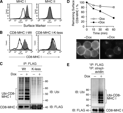
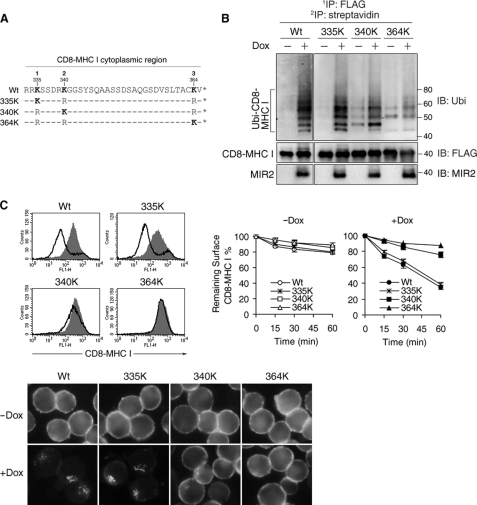
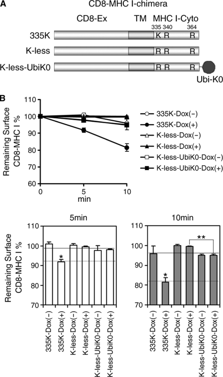
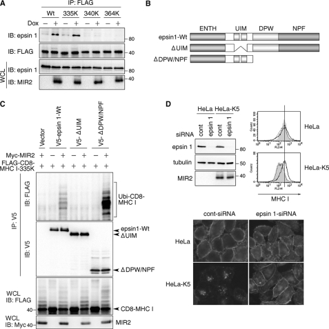
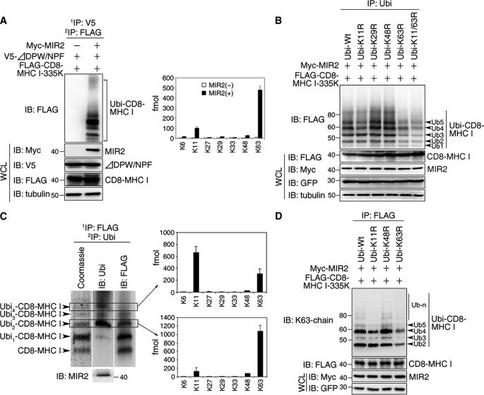
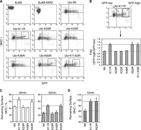
The polyubiquitin chain is generated by the sequential addition of ubiquitin moieties to target molecules, a reaction between specific lysine residues that is catalyzed by E3 ubiquitin ligase. The Lys(48)-linked and Lys(63)-linked polyubiquitin chains are well established inducers of proteasome-dependent degradation and signal transduction, respectively. The concept has recently emerged that polyubiquitin chain-mediated regulation is even more complex because various types of atypical polyubiquitin chains have been discovered in vivo. Here, we demonstrate that a novel complex ubiquitin chain functions as an internalization signal for major histocompatibility complex class I (MHC I) membrane proteins in vivo. Using a tetracycline-inducible expression system and quantitative mass spectrometry, we show that the polyubiquitin chain generated by the viral E3 ubiquitin ligase of Kaposi sarcoma-associated herpesvirus, MIR2, is a Lys(11) and Lys(63) mixed-linkage chain. This novel ubiquitin chain can function as an internalization signal for MHC I through its association with epsin1, an adaptor molecule containing ubiquitin-interacting motifs.


Figures

FIGURE 1.
FIGURE 1.
Ubiquitination is required for MHC I down-regulation in T-REx-MIR2 cells. A, after being incubated with or without Dox for 24 h, the expression level of MHC I and MHC II on T-REx-MIR2 cells was examined by flow cytometry. Data from the cells incubated with (open histograms) and without (shaded histograms) Dox are shown. B, the expression level of the indicated CD8 chimeras on T-REx-MIR2 cells was examined as in A. C, whole cell lysates extracted from the indicated CD8 chimera-expressing T-REx-MIR2 cells that had been cultivated with (+) or without (−) Dox for 6 h were incubated with an anti-FLAG Ab. Precipitated (IP) samples were probed (IB) with anti-FLAG M2 Ab (FLAG) or anti-ubiquitin FK2 Ab (Ubi). D, surface CD8-MHC I was labeled with anti-CD8 Ab after the cells were incubated with (+) or without (−) Dox for 6 h. After cultivation at 37 °C for the indicated times, the expression of the remaining surface CD8-MHC I was examined by staining with phycoerythrin-conjugated goat anti-mouse IgG. At each time point, the percentage of remaining CD8-MHC I was calculated relative to the value of labeled CD8-MHC I at 0 min (upper panel). In the lower panel, T-REx-MIR2 cells were incubated with Dox for 6 h, and FITC-conjugated anti-CD8 Ab was added for the last 10 min of culture. Internalized CD8-MHC I was observed by fluorescence microscopy. E, surface molecules on T-REx-MIR2 cells were biotinylated, and the cells were incubated with (+) or without (−) Dox for 6 h in the presence of 2 μm of bafilomycin A1. The biotinylated CD8-MHC I was sequentially purified with anti-FLAG Ab and streptavidin-agarose. Each sample was probed with FK2 Ab or M2 Ab.
FIGURE 2.
FIGURE 2.
Ubiquitination at Lys335 is a critical event for MHC I down-regulation by MIR2. A, protein sequences of the cytoplasmic region of each CD8 mutant chimera are aligned. B, the indicated CD8-mutant chimera-expressing T-REx-MIR2 cells were incubated with Dox for 6 h in the presence of 2 μm bafilomycin A1, and the ubiquitination status of the chimeras was examined as in Fig. 1E. C, after being incubated with or without Dox for 9 h, the expression level of the indicated CD8 chimeras on T-REx-MIR2 cells was examined by flow cytometry. Data from the cells incubated with (open histograms) and without (shaded histograms) Dox are shown (upper left). In the upper right panel, each surface-expressed CD8 chimera was labeled with anti-CD8 Ab after the cells were incubated with (+) or without (−) Dox for 6 h. After cultivation at 37 °C for the indicated times, the expression level of the CD8 chimeric proteins remaining on the cell surface was examined as in Fig. 1D. At each time point, the percentage of remaining CD8 chimera was calculated relative to its level at 0 min. In the lower panel, the indicated CD8 mutant chimera-expressing T-REx-MIR2 cells were incubated with Dox for 6 h, and FITC-conjugated anti-CD8 Ab was added for the last 10 min of culture. The internalized CD8 chimera was observed by fluorescence microscopy.
FIGURE 3.
FIGURE 3.
Polyubiquitination is necessary for efficient CD8-MHC I internalization. A, schematic representation of the CD8-chimera fusion proteins used in this study is shown. The extracellular region of CD8 (CD8-Ex) and the transmembrane (TM) and cytoplasmic regions of MHC I (MHC I-Cyto) are indicated. B, surface CD8 chimeras were labeled with anti-CD8 Ab after the cells were incubated with (+) or without (−) Dox for 24 h. After cultivation at 37 °C for the indicated times, the expression of the remaining surface CD8 chimera was examined as in Fig. 1D. At each time point, the percentage of remaining CD8 chimera was calculated relative to the value at 0 min (lower panel). The values are mean ± S.E. (error bars) (n = 3); *, p < 0.05 compared with 335K-Dox(−), K-less-Dox(− or +) and K-less-UbiK0-Dox(− or +). **, p < 0.05 K-less-UbiK0-Dox(+) was compared with K-less-Dox(+).
FIGURE 4.
FIGURE 4.
Epsin1 is an adaptor molecule for MIR2-mediated MHC I internalization. A, after incubation with or without Dox for 6 h, protein complexes that included the indicated CD8-MHC I chimeras were isolated from the indicated CD8-MHC I-expressing T-REx-MIR2 cells. The purified protein complexes were examined with anti-epsin1 Ab or M2 Ab. B, schematic representation of the epsin1 mutants used in this study is shown. The UIM, DPW domain, NPF domain, and epsin N-terminal homology domain (ENTH) are indicated. C, each indicated V5-tagged epsin1 was coexpressed with CD8-MHC I 335K and MIR2 in HeLa cells. Protein complexes that included each exogenous epsin1 were purified with anti-V5 Ab and analyzed by Western blotting with M2 and V5 Ab. The expression of CD8-MHC I 335K and MIR2 was examined in whole cell lysates with M2 Ab and anti-Myc Ab, respectively. D, control siRNA (scrambled) or siRNA for epsin1 was transfected into HeLa or MIR2-expressing HeLa cells. Whole cell lysates from the indicated cells were analyzed with anti-epsin1 and anti-tubulin Abs. The expression of MHC I in the indicated HeLa cells was examined by flow cytometry. The gray histogram indicates MHC I expression in nontransfected HeLa cells. The dotted line indicates the MHC I expression in control siRNA-transfected HeLa cells. The solid line indicates the MHC I expression in epsin1 siRNA-transfected HeLa cells. In the lower panel, the indicated transfected cells were cultivated with anti-MHC I Ab (W6/32) for 10 min. Internalized MHC I was observed by fluorescence microscopy as in Fig. 1D.
FIGURE 5.
FIGURE 5.
Generation of the Lys11 and Lys63 mixed-linkage ubiquitin chain at Lys335. A, CD8-MHC I 335K and V5-tagged epsin1 were coexpressed in HeLa cells with (MIR2(+)) or without MIR2 (MIR2(−)). First, the protein complex containing ubiquitinated CD8-MHC I 335K and epsin1 was purified with an anti-V5 Ab and denatured by boiling in 2% SDS buffer. The denatured, CD8-MHC I 335K was then purified with an anti-FLAG M2 Ab. After confirmation by Western blot analysis (left panel), each sample was analyzed by mass spectrometry (right panel). B, each indicated EGFP-tagged ubiquitin was coexpressed with CD8-MHC I 335K and MIR2 in HeLa cells. The ubiquitinated form of CD8-MHC I 335K was purified with the FK2 Ab and examined with the M2 Ab. Equal amounts of CD8-MHC I 335K were analyzed in all samples. C, the ubiquitinated forms of CD8-MHC I were sequentially purified from T-REx-MIR2 cells with M2 Ab and FK2 Ab and separated by SDS-PAGE. After confirmation by Western blot analysis with the indicated Abs, the peptides obtained from the bands indicated by the rectangle were analyzed by LC-MS/MS. D, each indicated EGFP-tagged ubiquitin was coexpressed with CD8-MHC I 335K and MIR2 in HeLa cells. The ubiquitinated form of CD8-MHC I 335K was purified with the M2 Ab and examined with the Lys63 linkage-specific Ab, Apu3. Equal amounts of CD8-MHC I 335K were analyzed in all samples.
FIGURE 6.
FIGURE 6.
Contribution of the Lys11 and Lys63 mixed-linkage ubiquitin chain to protein internalization. A, each indicated EGFP-tagged ubiquitin was expressed in constitutively MIR2-expressing BJAB cells. Two days after the introduction of each exogenous ubiquitin by electroporation, the expression level of MHC I was examined by flow cytometry. B, the surface expression levels of MHC I in low GFP-expressing and high GFP-expressing cell populations were measured as indicated in the upper panel, and the ratios of these two values are shown in the lower panel. The values are mean ± S.E. (error bars) (n = 3); *, p < 0.01 compared with wild-type ubiquitin (Wt). C, after each EGFP-tagged ubiquitin was expressed as in A, the down-regulation of surface MHC I at the indicated time points was examined in the cell population in which EGFP was highly expressed, as in Fig. 1D. The values are mean ± S.E. (n = 3); *, p < 0.01 compared with wild-type ubiquitin. D, the same experiment as in C was performed at a different time point (10 min). The values are mean ± S.E. (n = 3); *, p < 0.01 compared with wild-type ubiquitin.

Similar articles

Cited by

References

    1. Raiborg C., Stenmark H. (2009) Nature 458, 445–452 - PubMed
    1. Shtiegman K., Kochupurakkal B. S., Zwang Y., Pines G., Starr A., Vexler A., Citri A., Katz M., Lavi S., Ben-Basat Y., Benjamin S., Corso S., Gan J., Yosef R. B., Giordano S., Yarden Y. (2007) Oncogene 26, 6968–6978 - PubMed
    1. Gay D. L., Ramón H., Oliver P. M. (2008) Immunol. Res. 42, 51–64 - PMC - PubMed
    1. Gómez-Martín D., Díaz-Zamudio M., Alcocer-Varela J. (2008) Autoimmun. Rev. 7, 284–290 - PubMed
    1. Roepstorff K., Grøvdal L., Grandal M., Lerdrup M., van Deurs B. (2008) Histochem. Cell Biol. 129, 563–578 - PMC - PubMed

Publication types

MeSH terms