. 2010 Dec;299(6):L724-34.
doi: 10.1152/ajplung.00233.2010. Epub 2010 Sep 10.

Keratinocyte growth factor enhances barrier function without altering claudin expression in primary alveolar epithelial cells

Affiliations

Keratinocyte growth factor enhances barrier function without altering claudin expression in primary alveolar epithelial cells

Michael J LaFemina et al. Am J Physiol Lung Cell Mol Physiol. 2010 Dec.

Abstract

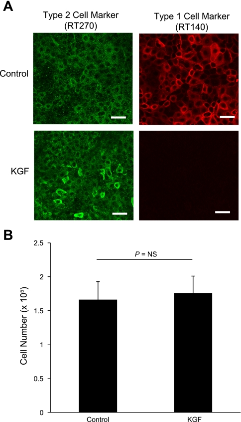
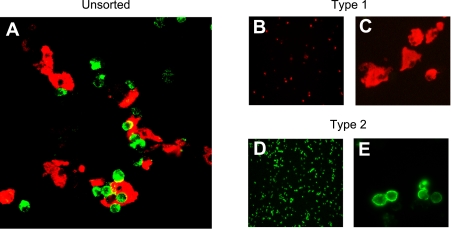
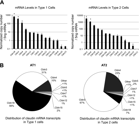
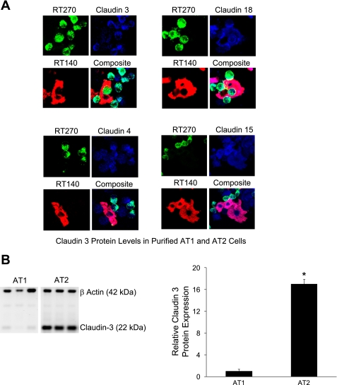
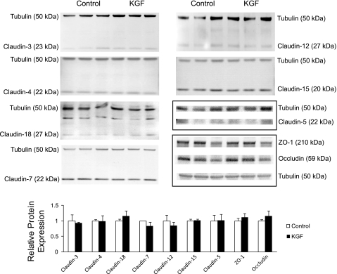
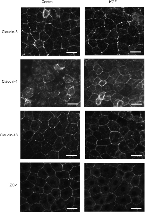
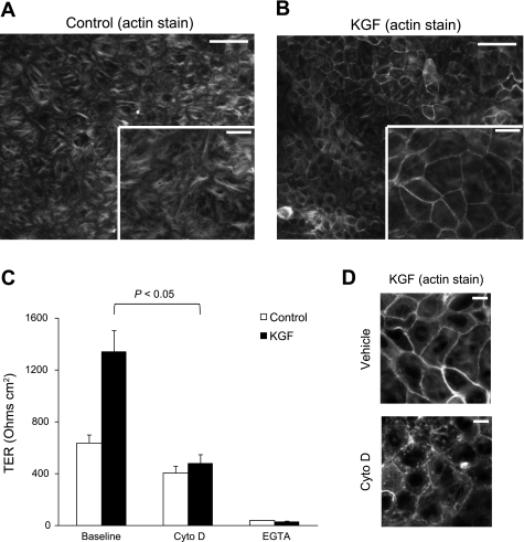
Keratinocyte growth factor (KGF) has efficacy in several experimental models of lung injury; however, the mechanisms underlying KGF's protective effect remain incompletely understood. This study was undertaken to determine whether KGF augments barrier function in primary rat alveolar epithelial cells grown in culture, specifically whether KGF alters tight junction function via claudin expression. KGF significantly increased alveolar epithelial barrier function in culture as assessed by transepithelial electrical resistance (TER) and paracellular permeability. Fluorescence-activated cell sorting of freshly isolated type 1 (AT1) and type 2 (AT2) cells followed by quantitative real-time RT-PCR revealed that more than 97% of claudin mRNA transcripts in these cells were for claudins-3, -4, and -18. Using cultured AT2 cells, we then examined the effect of KGF on the protein levels of the claudins with the highest mRNA levels: -3, -4, -5, -7, -12, -15, and -18. KGF did not alter the levels of any of the claudins tested, nor of zona occludens-1 (ZO-1) or occludin. Moreover, localization of claudins-3, -4, -18, and ZO-1 was unchanged. KGF did induce a marked increase in the apical perijunctional F-actin ring. Actin depolymerization with cytochalasin D blocked the KGF-mediated increase in TER without significantly changing TER in control cells. Together, these data support a novel mechanism by which KGF enhances alveolar barrier function, modulation of the actin cytoskeleton. In addition, these data demonstrate the complete claudin expression profile for AT1 and AT2 cells and indicate that claudins-3, -4, and -18 are the primary claudins expressed in these cell types.


Figures

Fig. 1.
Fig. 1.
Effect of keratinocyte growth factor (KGF) on barrier function of alveolar epithelial cells. Freshly isolated alveolar epithelial type 2 (AT2) cells were cultured for 5 days in control media or media supplemented with 10 ng/ml KGF. A: transepithelial electrical resistance (TER) was significantly greater in KGF-treated cells. *P < 0.001 compared with control, n = 9 biological replicates. B: apparent permeability (PAPP) to the 0.5 kDa tracer molecule pyranine was significantly decreased in KGF-treated cells. *P < 0.05 compared with control, n = 7 biological replicates. These values were less than 1% of PAPP across cell-free Transwells. Data are expressed as means ± SE.
Fig. 2.
Fig. 2.
Effect of KGF administration on alveolar epithelial cell phenotype and proliferation. Freshly isolated AT2 cells were cultured for 5 days in control media or media supplemented with KGF. A: at day 5, cells were fixed and immunolabeled for the AT2 marker RT270 (green) and the AT1 marker RT140 (red). Although cells expressed RT270 under both conditions, KGF-treated cells were more AT2-like given the absence of RT140 staining. Scale bars, 50 μm. B: cell number was measured in confluent monolayers of control and KGF-treated cells at day 5 in culture. There was no difference in cell number between the 2 groups as measured by quantification of crystal violet staining (n = 4 biological replicates). Data are expressed as means ± SE.
Fig. 3.
Fig. 3.
Isolation of AT1 and AT2 cells by fluorescence-activated cell sorting. Lung cells liberated by digestion with elastase were stained with anti-RT140 (AT1 cell marker, red) and anti-RT270 (AT2 cell marker, green) as described in text. A: cytocentrifuged preparation of mixed cell population before FACS analysis and sorting showing AT1 (red) and AT2 (green) cells together. B–E: cytocentrifuged preparations of essentially pure (>99%) AT1 (B and C, red) and AT2 (D and E) cells after FACS analysis and sorting. Magnification, ×10 (B and D) and ×40 (A, C, and E).
Fig. 4.
Fig. 4.
Claudin mRNA expression levels in freshly isolated, purified AT1 and AT2 cells. To determine which claudins are expressed in these cell types, mRNA expression was compared in AT1 and AT2 cells for all known claudins using quantitative real-time PCR as described in text. A: AT1 cells (left) expressed primarily claudins-18, -3, and -4 (*P < 0.05 compared with all other claudins expressed in AT1 cells) with detectable levels of claudins-15, -12, -7, -5, -19, -22, -11, -10b, -9, -20, and -23 in decreasing order. AT2 cells (right) expressed primarily claudins-3, -4, and -18, with detectable levels of -5, -7, -10b, -12, -19, -9, -15, -22, -20, and -23 in decreasing order (*P < 0.05 for claudins-3 and -4 compared with all other claudins expressed in AT2 cells). B: >97% of claudin mRNA transcripts in AT1 (left) and AT2 (right) cells were for claudins-3, -4, and -18, although the relative amounts of each of these varied with claudin-18 being most highly expressed in AT1 cells and claudin-3 being the dominant transcript in AT2 cells. After analysis of variance followed by Bonferroni correction for multiple comparisons, claudin-3 was the only transcript expressed at a statistically significantly different level between AT1 and AT2 cells with 10-fold less claudin-3 in AT1 cells (P < 0.05). Data are expressed as means ± SE.
Fig. 5.
Fig. 5.
Claudin protein expression in AT1 and AT2 cells. A: cytocentrifuged preparations of lung cells before FACS analysis and sorting were stained for RT140 (red, lower left), RT270 (green, top left), claudin-3, -4, -15, or -18 (all blue, top right). Coexpression of specific claudins with RT140 and RT270 was also assessed (bottom right). Claudin-3 was primarily expressed in AT2 cells, although staining in AT1 cells was also detected. Claudins-4, -15, and -18 were expressed in AT1 and AT2 cells. Of note, cells negative for both AT1 and AT2 markers in the unpurified cell preparations stained positive for claudins-15, -4, and -3. Magnification, ×40 for all images. B: immunoblot analysis of claudin-3 in isolated, purified AT1 and AT2 cells after flow cytometry. β-actin was also labeled to normalize for total sample protein content. Claudin-3 protein levels were 17-fold greater in AT2 cells (*P < 0.001, n = 3 biological replicates). Data are expressed as means ± SE.
Fig. 6.
Fig. 6.
Effect of KGF administration on the expression of tight junction proteins in alveolar epithelial cells. Freshly isolated AT2 cells were cultured on permeable supports for 5 days in control media or media supplemented with KGF and then harvested and resolved by SDS-PAGE. Levels of claudin-3, -4, -5, -7, -12, -15, -18, zonula occludens (ZO)-1, and occludin were determined by immunoblot. Immunoblots were also labeled for β-tubulin to normalize for total sample protein content. Densitometry revealed no significant differences in expression levels of these tight junction proteins in KGF-treated cells compared with control (n = 3 biological replicates). Data are expressed as means ± SE.
Fig. 7.
Fig. 7.
Localization of tight junction proteins cultured for 5 days in the absence or presence of KGF. Freshly isolated AT2 cells were plated on permeable supports in control media or media supplemented with KGF, and then cultured for 5 days, fixed, and immunostained for claudin-3, claudin-4, claudin-18, and ZO-1. There were no differences in localization noted for these tight junction proteins in KGF-treated cells compared with control. All proteins were predominantly localized to the membrane at cell-cell contacts. Scale bars, 20 μm.
Fig. 8.
Fig. 8.
Effect of KGF administration on the actin cytoskeleton. Freshly isolated AT2 cells were plated on permeable supports in control media or media supplemented with KGF, and then cultured for 5 days, fixed, stained for F-actin with Alexa 568 phalloidin, and imaged under epifluorescence (A and B) or confocal microscopy (D). A and B: KGF-treated cells (B) demonstrated significant reorganization of the actin cytoskeleton with a marked increase in the apical perijunctional F-actin ring compared with control cells (A). Inset is higher magnification image (n = at least 4 biological replicates). Scale bars, 50 μm on ×20 image and 20 μm on inset ×40 image. C: actin depolymerization with cytochalasin D treatment decreased TER to control levels (P < 0.05) without a significant decrease in control TER (n = 4 biological replicates consisting of 4–6 technical replicates). Treatment of control and KGF-treated AT2 cells with calcium-free media resulted in near complete loss of TER. Data are expressed as means ± SE. D: the perijunctional F-actin ring in KGF-treated alveolar epithelial cells (top) was disrupted by treatment with cytochalasin D (bottom) (n = at least 4 biological replicates). Scale bars, 10 μm.

Comment in

Similar articles

Cited by

References

    1. Abraham V, Chou ML, DeBolt KM, Koval M. Phenotypic control of gap junctional communication by cultured alveolar epithelial cells. Am J Physiol Lung Cell Mol Physiol 276: L825–L834, 1999 - PubMed
    1. Adamson IY, Bowden DH. The type 2 cell as progenitor of alveolar epithelial regeneration. A cytodynamic study in mice after exposure to oxygen. Lab Invest 30: 35–42, 1974 - PubMed
    1. Angelow S, Ahlstrom R, Yu AS. Biology of claudins. Am J Physiol Renal Physiol 295: F867–F876, 2008 - PMC - PubMed
    1. Atabai K, Ishigaki M, Geiser T, Ueki I, Matthay MA, Ware LB. Keratinocyte growth factor can enhance alveolar epithelial repair by nonmitogenic mechanisms. Am J Physiol Lung Cell Mol Physiol 283: L163–L169, 2002 - PubMed
    1. Baba Y, Yazawa T, Kanegae Y, Sakamoto S, Saito I, Morimura N, Goto T, Yamada Y, Kurahashi K. Keratinocyte growth factor gene transduction ameliorates acute lung injury and mortality in mice. Hum Gene Ther 18: 130–141, 2007 - PubMed

Publication types

MeSH terms