

Preexposure to hyperoxia causes increased lung injury and epithelial apoptosis in mice ventilated with high tidal volumes
- PMID: 20833778
- PMCID: PMC2980385
- DOI: 10.1152/ajplung.00072.2010
Preexposure to hyperoxia causes increased lung injury and epithelial apoptosis in mice ventilated with high tidal volumes
Abstract
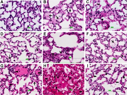
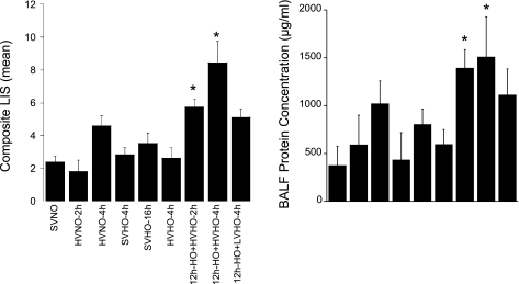
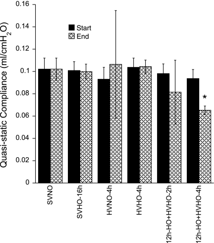
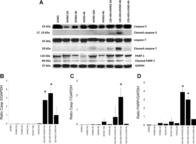
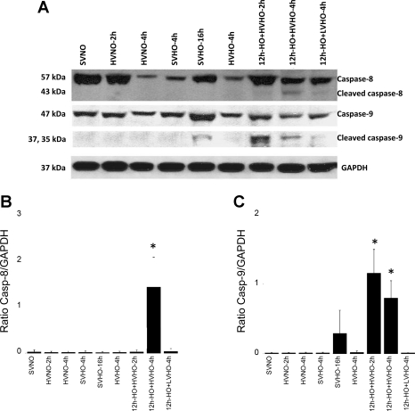
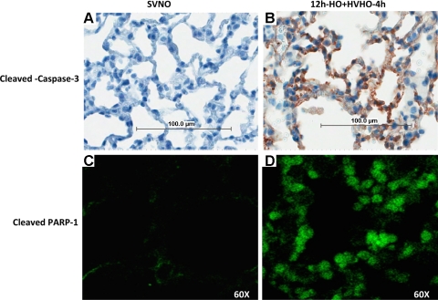
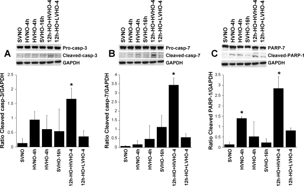
Both high tidal volume mechanical ventilation (HV) and hyperoxia (HO) have been implicated in ventilator-induced lung injury. However, patients with acute lung injury are often exposed to HO before the application of mechanical ventilation. The potential priming of the lungs for subsequent injury by exposure to HO has not been extensively studied. We provide evidence that HO (90%) for 12 h followed by HV (25 μl/g) combined with HO for 2 or 4 h (HO-12h+HVHO-2h or -4h) induced severe lung injury in mice. Analysis of lung homogenates showed that lung injury was associated with cleavage of executioner caspases, caspases-3 and -7, and their downstream substrate poly(ADP-ribose) polymerase-1 (PARP-1). No significant lung injury or caspase cleavage was seen with either HO for 16 h or HV for up to 4 h. Ventilation for 4 h with HO (HVHO) did not cause significant lung injury without preexposure to HO. Twelve-hour HO followed by lower tidal volume (6 μl/g) mechanical ventilation failed to produce significant injury or caspase cleavage. We also evaluated the initiator caspases, caspases-8 and -9, to determine whether the death receptor or mitochondrial-mediated pathways were involved. Caspase-9 cleavage was observed in HO-12h+HVHO-2h and -4h as well as HO for 16 h. Caspase-8 activation was observed only in HO-12h+HVHO-4h, indicating the involvement of both pathways. Immunohistochemistry and in vitro stretch studies showed caspase cleavage in alveolar epithelial cells. In conclusion, preexposure to HO followed by HV produced severe lung injury associated with alveolar epithelial cell apoptosis.
Figures
Similar articles
-
Lung injury caused by high tidal volume mechanical ventilation and hyperoxia is dependent on oxidant-mediated c-Jun NH2-terminal kinase activation.J Appl Physiol (1985). 2011 Nov;111(5):1467-76. doi: 10.1152/japplphysiol.00539.2011. Epub 2011 Jul 28. J Appl Physiol (1985). 2011. PMID: 21799126 Free PMC article.
-
Poly(ADP-ribose)polymerase activation mediates lung epithelial cell death in vitro but is not essential in hyperoxia-induced lung injury.Am J Respir Cell Mol Biol. 2005 Dec;33(6):555-64. doi: 10.1165/rcmb.2004-0361OC. Epub 2005 Sep 8. Am J Respir Cell Mol Biol. 2005. PMID: 16151053
-
Activation of Src-dependent Smad3 signaling mediates the neutrophilic inflammation and oxidative stress in hyperoxia-augmented ventilator-induced lung injury.Respir Res. 2015 Sep 16;16(1):112. doi: 10.1186/s12931-015-0275-6. Respir Res. 2015. PMID: 26377087 Free PMC article.
-
Lung Injury After One-Lung Ventilation: A Review of the Pathophysiologic Mechanisms Affecting the Ventilated and the Collapsed Lung.Anesth Analg. 2015 Aug;121(2):302-18. doi: 10.1213/ANE.0000000000000808. Anesth Analg. 2015. PMID: 26197368 Review.
-
Ventilator-induced lung injury: the anatomical and physiological framework.Crit Care Med. 2010 Oct;38(10 Suppl):S539-48. doi: 10.1097/CCM.0b013e3181f1fcf7. Crit Care Med. 2010. PMID: 21164395 Review.
Cited by
-
CXCR4 regulates migration of lung alveolar epithelial cells through activation of Rac1 and matrix metalloproteinase-2.Am J Physiol Lung Cell Mol Physiol. 2012 May 1;302(9):L846-56. doi: 10.1152/ajplung.00321.2011. Epub 2012 Feb 17. Am J Physiol Lung Cell Mol Physiol. 2012. PMID: 22345572 Free PMC article.
-
Deletion of apoptosis signal-regulating kinase-1 prevents ventilator-induced lung injury in mice.Am J Respir Cell Mol Biol. 2012 Apr;46(4):461-9. doi: 10.1165/rcmb.2011-0234OC. Epub 2011 Nov 3. Am J Respir Cell Mol Biol. 2012. PMID: 22052879 Free PMC article.
-
Mechanical Ventilation Exacerbates Poly (I:C) Induced Acute Lung Injury: Central Role for Caspase-11 and Gut-Lung Axis.Front Immunol. 2021 Jul 19;12:693874. doi: 10.3389/fimmu.2021.693874. eCollection 2021. Front Immunol. 2021. PMID: 34349759 Free PMC article.
-
Regulation of interleukin-6 secretion by the two-pore-domain potassium channel Trek-1 in alveolar epithelial cells.Am J Physiol Lung Cell Mol Physiol. 2013 Feb 15;304(4):L276-86. doi: 10.1152/ajplung.00299.2012. Epub 2012 Dec 28. Am J Physiol Lung Cell Mol Physiol. 2013. PMID: 23275623 Free PMC article.
-
Impact of hyperoxemia on mortality in critically ill patients with ventilator-associated pneumonia.Ann Transl Med. 2018 Nov;6(21):417. doi: 10.21037/atm.2018.10.19. Ann Transl Med. 2018. PMID: 30581825 Free PMC article.
References
-
- The Acute Respiratory Distress Syndrome Network Ventilation with lower tidal volumes as compared with traditional tidal volumes for acute lung injury and the acute respiratory distress syndrome. The Acute Respiratory Distress Syndrome Network. N Engl J Med 342: 1301–1308, 2000 - PubMed
-
- Altemeier WA, Sinclair SE. Hyperoxia in the intensive care unit: why more is not always better. Curr Opin Crit Care 13: 73–78, 2007 - PubMed
-
- Bailey TC, Martin EL, Zhao L, Veldhuizen RA. High oxygen concentrations predispose mouse lungs to the deleterious effects of high stretch ventilation. J Appl Physiol 94: 975–982, 2003 - PubMed
-
- Clerch LB, Baras AS, Massaro GD, Hoffman EP, Massaro D. DNA microarray analysis of neonatal mouse lung connects regulation of KDR with dexamethasone-induced inhibition of alveolar formation. Am J Physiol Lung Cell Mol Physiol 286: L411–L419, 2004 - PubMed
-
- Davis JM, Penney DP, Notter RH, Metlay L, Dickerson B, Shapiro DL. Lung injury in the neonatal piglet caused by hyperoxia and mechanical ventilation. J Appl Physiol 67: 1007–1012, 1989 - PubMed
Publication types
MeSH terms
Substances
Grants and funding
LinkOut - more resources
Full Text Sources
Other Literature Sources
Miscellaneous