

Interaction of Bacillus thuringiensis vegetative insecticidal protein with ribosomal S2 protein triggers larvicidal activity in Spodoptera frugiperda
- PMID: 20833785
- PMCID: PMC2976263
- DOI: 10.1128/AEM.01552-10
Interaction of Bacillus thuringiensis vegetative insecticidal protein with ribosomal S2 protein triggers larvicidal activity in Spodoptera frugiperda
Abstract
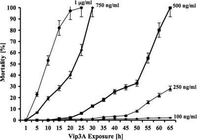
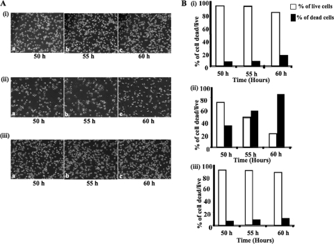
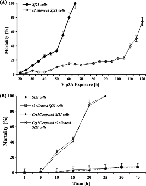
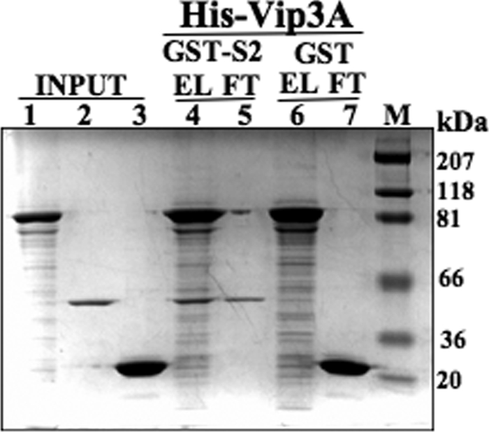
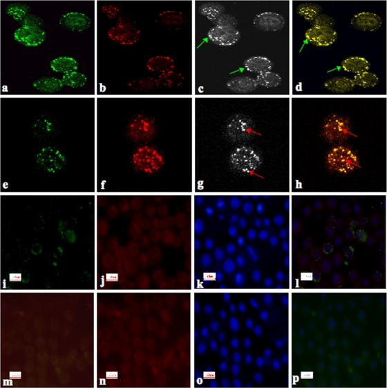
Vegetative insecticidal protein (Vip3A) is synthesized as an extracellular insecticidal toxin by certain strains of Bacillus thuringiensis. Vip3A is active against several lepidopteran pests of crops. Polyphagous pest, Spodoptera frugiperda, and its cell line Sf21 are sensitive for lyses to Vip3A. Screening of cDNA library prepared from Sf21 cells through yeast two-hybrid system with Vip3A as bait identified ribosomal protein S2 as a toxicity-mediating interacting partner protein. The Vip3A-ribosomal-S2 protein interaction was validated by in vitro pulldown assays and by RNA interference-induced knockdown experiments. Knockdown of expression of S2 protein in Sf21 cells resulted in reduced toxicity of the Vip3A protein. These observations were further extended to adult fifth-instar larvae of Spodoptera litura. Knockdown of S2 expression by injecting corresponding double-stranded RNA resulted in reduced mortality of larvae to Vip3A toxin. Intracellular visualization of S2 protein and Vip3A through confocal microscopy revealed their interaction and localization in cytoplasm and surface of Sf21 cells.
Figures
Similar articles
-
[Screening of Bacillus thuringiensis strains containing vip3A genes and analysis of gene conservation].Sheng Wu Gong Cheng Xue Bao. 2003 Sep;19(5):538-44. Sheng Wu Gong Cheng Xue Bao. 2003. PMID: 15969080 Chinese.
-
Vip3A, a novel Bacillus thuringiensis vegetative insecticidal protein with a wide spectrum of activities against lepidopteran insects.Proc Natl Acad Sci U S A. 1996 May 28;93(11):5389-94. doi: 10.1073/pnas.93.11.5389. Proc Natl Acad Sci U S A. 1996. PMID: 8643585 Free PMC article.
-
[Effects of helper protein P20 from Bacillus thuringiensis on Vip3A expression].Wei Sheng Wu Xue Bao. 2006 Feb;46(1):85-9. Wei Sheng Wu Xue Bao. 2006. PMID: 16579471 Chinese.
-
Bacillus thuringiensis vegetative insecticidal protein family Vip3A and mode of action against pest Lepidoptera.Pest Manag Sci. 2020 May;76(5):1612-1617. doi: 10.1002/ps.5804. Epub 2020 Mar 11. Pest Manag Sci. 2020. PMID: 32103608 Review.
-
Managing the Invasive Fall Armyworm through Biotech Crops: A Chinese Perspective.Trends Biotechnol. 2021 Feb;39(2):105-107. doi: 10.1016/j.tibtech.2020.07.001. Epub 2020 Jul 23. Trends Biotechnol. 2021. PMID: 32713608 Review.
Cited by
-
Reduced Membrane-Bound Alkaline Phosphatase Does Not Affect Binding of Vip3Aa in a Heliothis virescens Resistant Colony.Toxins (Basel). 2020 Jun 19;12(6):409. doi: 10.3390/toxins12060409. Toxins (Basel). 2020. PMID: 32575644 Free PMC article.
-
Toxicity of Cry- and Vip3Aa-Class Proteins and Their Interactions against Spodoptera frugiperda (Lepidoptera: Noctuidae).Toxins (Basel). 2024 Apr 16;16(4):193. doi: 10.3390/toxins16040193. Toxins (Basel). 2024. PMID: 38668618 Free PMC article.
-
Cross-pollination in seed-blended refuge and selection for Vip3A resistance in a lepidopteran pest as detected by genomic monitoring.Proc Natl Acad Sci U S A. 2024 Mar 26;121(13):e2319838121. doi: 10.1073/pnas.2319838121. Epub 2024 Mar 21. Proc Natl Acad Sci U S A. 2024. PMID: 38513093 Free PMC article.
-
Structural and Functional Insights into the C-terminal Fragment of Insecticidal Vip3A Toxin of Bacillus thuringiensis.Toxins (Basel). 2020 Jul 5;12(7):438. doi: 10.3390/toxins12070438. Toxins (Basel). 2020. PMID: 32635593 Free PMC article.
-
Dissimilar Regulation of Antimicrobial Proteins in the Midgut of Spodoptera exigua Larvae Challenged with Bacillus thuringiensis Toxins or Baculovirus.PLoS One. 2015 May 18;10(5):e0125991. doi: 10.1371/journal.pone.0125991. eCollection 2015. PLoS One. 2015. PMID: 25993013 Free PMC article.
References
-
- Arora, N., A. Selvapandiyan, N. Agrawal, and R. K. Bhatnagar. 2003. Relocating expression of vegetative insecticidal protein into mother cell of Bacillus thuringiensis. Biochem. Biophys. Res. Commun. 310:158-162. - PubMed
-
- Beckmann, G., and P. Bork. 1993. An adhesive domain detected in functionally diverse receptors. Trends Biochem. Sci. 18:40-41. - PubMed
-
- Bradford, M. M. 1976. A rapid and sensitive method for the quantitation of microgram quantities of protein utilizing the principle of protein-dye binding. Anal. Biochem. 72:248-254. - PubMed
-
- Cioffi, M., and M. G. Wolfersberger. 1983. Isolation of separate apical, lateral, and basal plasma membrane from cells of an insect epithelium: a procedure based on tissue organization and ultrastructure. Tissue Cell 15:781-803. - PubMed
Publication types
MeSH terms
Substances
Grants and funding
LinkOut - more resources
Full Text Sources
Other Literature Sources
