. 2010 Nov;192(21):5767-77.
doi: 10.1128/JB.00624-10. Epub 2010 Sep 10.

Role of cross talk in regulating the dynamic expression of the flagellar Salmonella pathogenicity island 1 and type 1 fimbrial genes

Affiliations

Role of cross talk in regulating the dynamic expression of the flagellar Salmonella pathogenicity island 1 and type 1 fimbrial genes

Supreet Saini et al. J Bacteriol. 2010 Nov.

Abstract

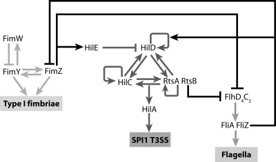
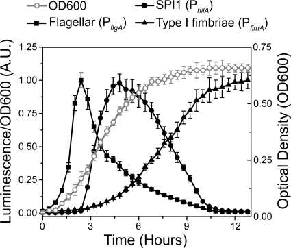
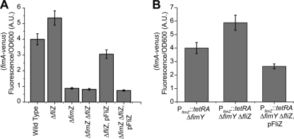
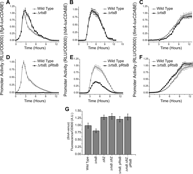
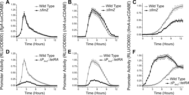
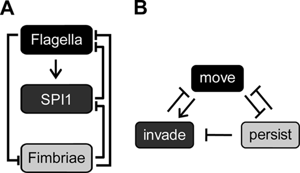
Salmonella enterica, a common food-borne pathogen, differentially regulates the expression of multiple genes during the infection cycle. These genes encode systems related to motility, adhesion, invasion, and intestinal persistence. Key among them is a type three secretion system (T3SS) encoded within Salmonella pathogenicity island 1 (SPI1). In addition to the SPI1 T3SS, other systems, including flagella and type 1 fimbriae, have been implicated in Salmonella pathogenesis. In this study, we investigated the dynamic expression of the flagellar, SPI1, and type 1 fimbrial genes. We demonstrate that these genes are expressed in a temporal hierarchy, beginning with the flagellar genes, followed by the SPI1 genes, and ending with the type 1 fimbrial genes. This hierarchy could mirror the roles of these three systems during the infection cycle. As multiple studies have shown that extensive regulatory cross talk exists between these three systems, we also tested how removing different regulatory links between them affects gene expression dynamics. These results indicate that cross talk is critical for regulating gene expression during transitional phases in the gene expression hierarchy. In addition, we identified a novel regulatory link between flagellar and type 1 fimbrial gene expression dynamics, where we found that the flagellar regulator, FliZ, represses type 1 fimbrial gene expression through the posttranscriptional regulation of FimZ. The significance of these results is that they provide the first systematic study of the effect of regulatory cross talk on the expression dynamics of flagellar, SPI1, and type 1 fimbrial genes.


Figures

FIG. 1.
FIG. 1.
Coordinate regulation of the flagellar, SPI1, and type 1 fimbrial genes. The master regulator for flagellar gene expression is FlhD4C2 (9). The FlhD4C2 complex, in turn, activates two additional regulators, FliA and FliZ, encoded within the fliAZ operon. FliA is a flagellum-specific alternate sigma factor essential for the expression of the motor, filament, and chemotaxis genes. FliZ is a posttranslational activator of FlhD4C2 (71). FliZ also activates HilD (38, 41, 53, 57, 82) and represses FimZ posttranscriptionally (this study). In the SPI1 T3SS, HilD, HilC, and RtsA form three interlocking positive-feedback loops where all three activate each other's and their own expression (23). In addition, they can independently activate HilA expression. HilA is required for the expression of the genes encoding the SPI1 T3SS. SPI1 gene expression is negatively regulated by HilE, which binds to HilD and prevents it from activating the SPI1 promoters. RtsB, encoded within the same operon as RtsA, binds to the PflhDC promoter and represses motility (22). In type 1 fimbriae, FimW and FimZ form a coupled feedback loop where they can activate their own and each other's expression (72). They can also independently activate the expression of the PfimA promoter, which controls the expression of genes encoding type 1 fimbriae. FimY and FimW also participate in a negative-feedback loop, where FimY activates FimW expression and FimW represses FimY expression. FimZ also binds to the PflhDC promoter and represses the expression of the flagellar genes (10) and induces the expression of HilE to repress SPI1 gene expression (5, 72).
FIG. 2.
FIG. 2.
Dynamic expression of the flagellar, SPI1, and type 1 fimbrial genes. Time course dynamics of the PflgA (flagellar), PhilA (SPI1), and PfimA (type 1 fimbrial) promoter activities in wild-type cells as determined using luciferase transcriptional reporters. For reference purposes, the optical density (OD600) was plotted to illustrate how each system is activated during different phases of growth. In these experiments, cells were first grown overnight at 37°C in LB without salt and then subcultured 1:500 in fresh LB-1% salt. Cells were then grown statically with luminescence, and optical density readings were taken every 20 min. Average promoter activities from three independent experiments on separate days are reported. For each experiment, six samples were tested. Error bars denote standard deviations. A.U., arbitrary units.
FIG. 3.
FIG. 3.
Effect of transcriptional cross talk on flagellar, SPI1, and type 1 fimbrial gene expression dynamics. (A to C) The flagellar genes amplify SPI1 gene expression and delay type 1 fimbrial gene expression. PflgA (A), PhilA (B), and PfimA (C) promoter activities in the wild type and the ΔflhDC mutant are shown. (D to F) The SPI1 genes reduce the duration of flagellar gene expression and accelerate the induction of type 1 fimbrial gene expression. PflgA (D), PhilA (E), and PfimA (F) promoter activities in the wild type and the ΔSPI1 mutant are shown. (G to I) Type 1 fimbrial genes do not affect flagellar gene expression and reduce the duration of SPI1 gene expression. PflgA (G), PhilA (H), and PfimA (I) promoter activities in the wild type and the ΔfimYZW mutant are shown. Experiments were performed as described for Fig. 2. The mutants were also tested to see whether they affected growth. However, no change in optical density as a function of time was observed (data not shown).
FIG. 4.
FIG. 4.
FliZ controls the magnitude of SPI1 gene expression and the dynamics of type 1 fimbrial gene expression. (A to C) Deleting FliZ represses flagellar and SPI1 gene expression and accelerates the induction of type 1 fimbrial genes. PflgA (A), PhilA (B), and PfimA (C) promoter activities in the wild type and the ΔfliZ mutant are shown. (D to F) Overexpressing FliZ increases the magnitude of flagellar and SPI1 gene expression and delays the induction of type 1 fimbrial genes. PflgA (D), PhilA (E), and PfimA (F) promoter activities in the wild type and the ΔfliZ mutant constitutively expressing FliZ from a PLtetO-1 promoter on a plasmid (pFliZ) are shown. Experiments were performed as described for Fig. 2.
FIG. 5.
FIG. 5.
FliZ regulates type 1 fimbrial gene expression though FimZ. (A) FliZ is unable to regulate PfimA promoter activity in the absence of FimZ. PfimA promoter activities in the wild type, the ΔfimZ mutant, the ΔfliZ mutant, the ΔfimZ ΔfliZ mutant, the ΔfliZ mutant expressing FliZ from the constitutive PLtetO-1 promoter on a plasmid (pFliZ), and the ΔfimZ ΔfliZ mutant harboring pFliZ are shown. (B) FliZ regulates FimZ posttranscriptionally. PfimA promoter activities in the PfimZ::tetRA ΔfimY mutant, the PfimZ::tetRA ΔfimY ΔfliZ mutant, and the PfimZ::tetRA ΔfimY ΔfliZ mutant expressing FliZ from the constitutive PLtetO-1 promoter on a plasmid are shown. In the genetic background PfimZ::tetRA, FimZ is under the control of a tetracycline-inducible promoter. Overnight cultures were subcultured 1:1,000 in fresh LB and then grown statically at 37°C for 24 h. FimZ expression was induced by addition of 15 μg/ml tetracycline. Fluorescence and optical density (OD600) values were then measured for each sample. Average promoter activities from three independent experiments on separate days are reported. For each experiment, six samples were tested. Error bars denote standard deviations.
FIG. 6.
FIG. 6.
RtsB controls the dynamics of flagellar and type 1 fimbrial gene expression. (A to C) Deleting RtsB increases the duration of flagellar gene expression and slows the induction of type 1 fimbrial genes. PflgA (A), PhilA (B), and PfimA (C) promoter activities in the wild type and the ΔrtsB mutant are shown. (D to F) Overexpressing RtsB inactivates flagellar gene expression and accelerates the induction of type 1 fimbrial genes. PflgA (D), PhilA (E), and PfimA (F) promoter activities in the wild type and the ΔrtsB mutant constitutively expressing RtsB from the constitutive PLtetO-1 promoter on a plasmid (pRtsB) are shown. Dynamic luminescence experiments were performed as described for Fig. 2. (G) RtsB regulates type 1 fimbrial gene expression through FliZ. PfimA promoter activities in the wild type, the ΔrtsB mutant, the ΔfliZ mutant, the ΔfliZ ΔrtsB mutant, and the ΔrtsB and ΔfliZ ΔrtsB mutants constitutively expressing RtsB are shown. Endpoint fluorescence experiments were performed as described for Fig. 5.
FIG. 7.
FIG. 7.
FimZ controls the dynamics of SPI1 gene expression. (A to C) Deleting FimZ increases the duration of SPI1 gene expression. PflgA (A), PhilA (B), and PfimA (C) promoter activities in the wild type and the ΔfimZ mutant are shown. (D to F) Overexpressing FimZ represses both flagellar and SPI1 gene expression. PflgA (D), PhilA (E), and PfimA (F) promoter activities in the wild type and the PfimZ::tetRA mutant, where FimZ is under the control of a tetracycline-inducible promoter, are shown. FimZ expression was induced by addition of 15 μg/ml tetracycline. Experiments were performed as described for Fig. 2.
FIG. 8.
FIG. 8.
Salmonella invasion program. (A) Diagram of transcriptional cross talk between the flagellar, SPI1, and type 1 fimbrial gene systems. (B) Inferred logic of transcriptional cross talk, where the decision to “move” results from flagellar gene expression, the decision to “invade” results from SPI1 gene expression, and the decision to “persist” results from type 1 fimbrial gene expression.

References

    1. Althouse, C., S. Patterson, P. Fedorka-Cray, and R. E. Isaacson. 2003. Type 1 fimbriae of Salmonella enterica serovar Typhimurium bind to enterocytes and contribute to colonization of swine in vivo. Infect. Immun. 71:6446-6452. - PMC - PubMed
    1. Aslanzadeh, J., and L. J. Paulissen. 1992. Role of type 1 and type 3 fimbriae on the adherence and pathogenesis of Salmonella enteritidis in mice. Microbiol. Immunol. 36:351-359. - PubMed
    1. Bajaj, V., R. L. Lucas, C. Hwang, and C. A. Lee. 1996. Co-ordinate regulation of Salmonella typhimurium invasion genes by environmental and regulatory factors is mediated by control of hilA expression. Mol. Microbiol. 22:703-714. - PubMed
    1. Baumler, A. J., R. M. Tsolis, and F. Heffron. 1996. Contribution of fimbrial operons to attachment to and invasion of epithelial cell lines by Salmonella typhimurium. Infect. Immun. 64:1862-1865. - PMC - PubMed
    1. Baxter, M. A., and B. D. Jones. 2005. The fimYZ genes regulate Salmonella enterica serovar Typhimurium invasion in addition to type 1 fimbrial expression and bacterial motility. Infect. Immun. 73:1377-1385. - PMC - PubMed

Publication types

MeSH terms

LinkOut - more resources