. 2010 Dec;9(12):1925-35.
doi: 10.1128/EC.00022-10. Epub 2010 Sep 10.

Schizosaccharomyces pombe calmodulin, Cam1, plays a crucial role in sporulation by recruiting and stabilizing the spindle pole body components responsible for assembly of the forespore membrane

Affiliations

Schizosaccharomyces pombe calmodulin, Cam1, plays a crucial role in sporulation by recruiting and stabilizing the spindle pole body components responsible for assembly of the forespore membrane

Akiko Itadani et al. Eukaryot Cell. 2010 Dec.

Abstract

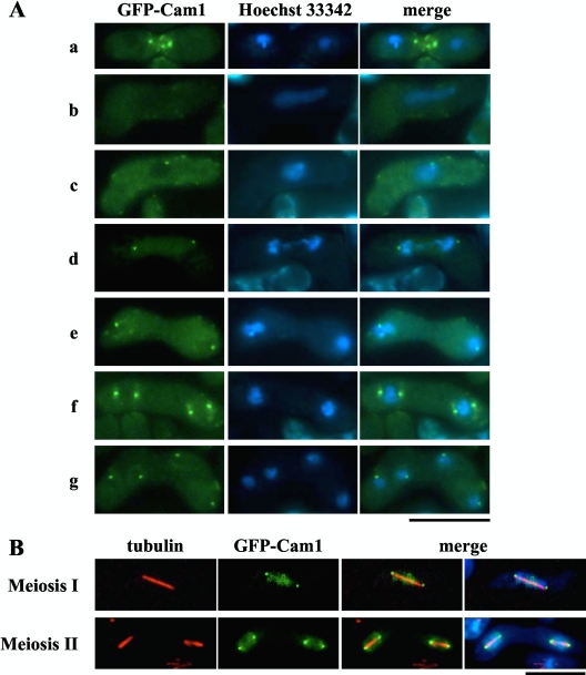
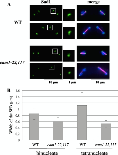
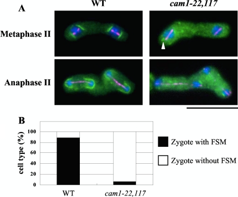
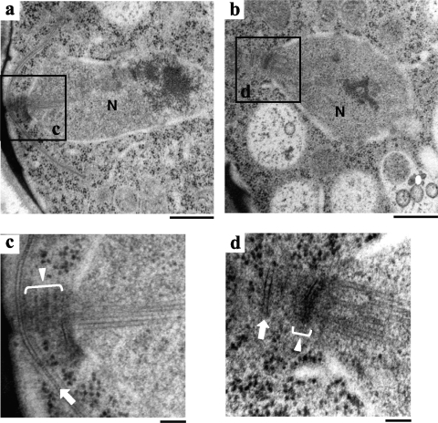
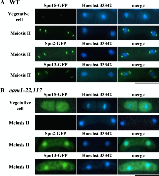
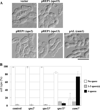
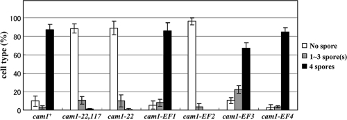
Calmodulin in Schizosaccharomyces pombe is encoded by the cam1(+) gene, which is indispensable for both vegetative growth and sporulation. Here, we report how Cam1 functions in spore formation. We found that Cam1 preferentially localized to the spindle pole body (SPB) during meiosis and sporulation. Formation of the forespore membrane, a precursor of the plasma membrane in spores, was blocked in a missense cam1 mutant, which was viable but unable to sporulate. Three SPB proteins necessary for the onset of forespore membrane formation, Spo2, Spo13, and Spo15, were unable to localize to the SPB in the cam1 mutant although five core SPB components that were tested were present. Recruitment of Spo2 and Spo13 is known to require the presence of Spo15 in the SPB. Notably, Spo15 was unstable in the cam1 mutant, and as a result, SPB localization of Spo2 and Spo13 was lost. Overexpression of Spo15 partially alleviated the sporulation defect in the cam1 mutant. These results indicate that calmodulin plays an essential role in forespore membrane formation by stably maintaining Spo15, and thus Spo2 and Spo13, at the SPB in meiotic cells.


Figures

Fig. 1.
Fig. 1.
Cellular localization of Cam1 during mating, meiosis, and sporulation. The homothallic haploid strain AI248 harboring a gfp-cam1 fusion gene was cultured on SSA medium to induce the sexual cycle. (A) Cells were stained with Hoechst 33342 to visualize nuclei. Developmental stages shown are as follows: row a, before karyogamy; row b, horsetail stage; row c, before meiosis I; row d, meiosis I; row e, interphase; row f, early meiosis II; row g, after meiosis II. Bar, 10 μm. (B) Cells were fixed during meiosis. Microtubules and nuclei were visualized by anti α-tubulin antibody, TAT1 (red) (32), and DAPI (blue), respectively. Bar, 10 μm.
Fig. 2.
Fig. 2.
Structural modification of the SPB during sporulation. The TN8 (h90 cam1+) and AI22 (h90 cam1-22,117) strains were cultured on MEA sporulation medium. (A) Immunostaining of the SPB with anti-Sad1 antibody (green). Magnified images of the boxed regions are also presented. Meiotic stages of the cells were examined by staining nuclei with DAPI (blue) and spindle microtubules with anti-α-tubulin antibody, TAT1 (magenta). (B) Comparison of the width of the SPB between wild-type and cam1-22,117 strains. Binucleate and tetranucleate cells were determined separately. The means ± standard deviations of approximately 100 SPBs are shown.
Fig. 3.
Fig. 3.
Initiation of FSM assembly in the cam1-22,117 mutant. (A) The nascent FSM was visualized by GFP-psy1 (green). A cam1+ strain (YM20) and a cam1-22,117 strain (AI52) were cultured on MEA sporulation medium and doubly stained with anti-α-tubulin antibody, TAT1 (magenta), and DAPI (blue). The arrowhead indicates a very short FSM-like structure. Bar, 10 μm. (B) Quantitative assay for zygote formation with an FSM. Only zygotes that assembled meiosis II spindles were counted.
Fig. 4.
Fig. 4.
Fine structures of wild-type and cam1-22,117 strains during meiosis II. Cells were fixed after incubation on MEA sporulation medium for 1 day. Electron microscopic images of the wild-type (a and c) and cam1-22,117 (b and d) strains. Panels c and d are magnified images of the boxed regions in panels a and b, respectively. N, nucleus; white lines with triangles, the SPB; white arrows, the expanding FSM (c) and the abortive FSM (d). Bars, 500 nm (a and b) and 100 nm (c and d).
Fig. 5.
Fig. 5.
Localization of Spo15, Spo2, and Spo13 in wild-type and cam1-22,117 strains. (A) Homothallic wild-type (cam1+) strains expressing Spo15-GFP (AI154), Spo2-GFP (AI259), or Spo13-GFP (AI258) were cultured on complete (YEA) and sporulation (SSA) media. Cells were stained with Hoechst 33342 (blue). Bar, 10 μm. (B) Similarly, homothallic cam1-22,117 strains, expressing Spo15-GFP (AI156), Spo2-GFP (AI262), or Spo13-GFP (AI263), were cultured on YEA and SSA media. Bar, 10 μm.
Fig. 6.
Fig. 6.
Detection of Spo15 and Cam1 by Western blotting. (A) Western analysis of Spo15. Strains AI510 (cam1+), AI512 (cam1-22), and AI518 (cam1-22,117) were cultured in liquid synthetic medium, MM+N, for 20 h (vegetative growth) and liquid sporulation medium, MM-N, for 8 h (sporulation). Protein extracts were subjected to immunoblotting with anti-Spo15 antibody and with anti-α-tubulin antibody as a loading control. (B) Western analysis of Cam1. AI248 (cam1+) and AI210 (spo15Δ) strains expressing GFP-Cam1 were cultured in MM+N for 8 h and in MM-N for 6 h. Protein extracts were subjected to immunoblotting with anti-GFP antibody and with anti-α-tubulin antibody as a loading control. WT, wild type.
Fig. 7.
Fig. 7.
Overexpression of Spo15, Spo2, and Spo13 in the cam1-22,117 mutant. (A) The AI22 (cam1-22,117) strain was transformed with a pREP1 expression vector carrying spo15+ spo2+ or spo13+ and a multicopy plasmid carrying cam1+, pAL(cam1). Transformants were cultured on sporulation medium (SSA) for 2 days. Bar, 10 μm. (B) Sporulation was monitored by determining the percentage of asci, which were further classified according to the number of spores per ascus.
Fig. 8.
Fig. 8.
Sporulation ability of the cam1 mutant harboring mutations in EF-hand motifs. The wild-type cam1+ and respective cam1 mutant genes were integrated at the leu1 locus on chromosome II of AI509. Strains used were AI510 (cam1+), AI512 (cam1-22), AI518 (cam1-22, 117), AI520 (cam1-EF1), AI522 (cam1-EF2), AI524 (cam1-EF3), and AI526 (cam1-EF4). Strains were cultured on MEA medium for 2 days, and their sporulation efficiencies were measured. The percentage of asci, classified according to the number of spores per ascus, is presented.

Similar articles

Cited by

References

    1. Aalto M. K., Ronne H., Keränen S. 1993. Yeast syntaxins Sso1p and Sso2p belong to a family of related membrane proteins that function in vesicular transport. EMBO J. 12:4095–4104 - PMC - PubMed
    1. Bennett M. K., Calakos N., Scheller R. H. 1992. Syntaxin: a synaptic protein implicated in docking of synaptic vesicles at presynaptic active zones. Science 257:255–259 - PubMed
    1. Chikashige Y., Ding D. Q., Funabiki H., Mashiko S., Yanagida M., Hiraoka Y. 1994. Telomere-led premeiotic chromosome movement in fission yeast. Science 264:270–273 - PubMed
    1. Cyert M. S. 2001. Genetic analysis of calmodulin and its targets in Saccharomyces cerevisiae. Annu. Rev. Genet. 35:647–672 - PubMed
    1. Davis T. N., Urdea M. S., Masiarz F. R., Thorner J. 1986. Isolation of the yeast calmodulin gene: calmodulin is an essential protein. Cell 47:423–431 - PubMed

Publication types

MeSH terms

Substances

LinkOut - more resources