. 2010 Nov 25;116(22):4646-56.
doi: 10.1182/blood-2010-04-280925. Epub 2010 Sep 10.

Transcription profiling in human platelets reveals LRRFIP1 as a novel protein regulating platelet function

Affiliations

Transcription profiling in human platelets reveals LRRFIP1 as a novel protein regulating platelet function

Alison H Goodall et al. Blood. .

Abstract

Within the healthy population, there is substantial, heritable, and interindividual variability in the platelet response. We explored whether a proportion of this variability could be accounted for by interindividual variation in gene expression. Through a correlative analysis of genome-wide platelet RNA expression data from 37 subjects representing the normal range of platelet responsiveness within a cohort of 500 subjects, we identified 63 genes in which transcript levels correlated with variation in the platelet response to adenosine diphosphate and/or the collagen-mimetic peptide, cross-linked collagen-related peptide. Many of these encode proteins with no reported function in platelets. An association study of 6 of the 63 genes in 4235 cases and 6379 controls showed a putative association with myocardial infarction for COMMD7 (COMM domain-containing protein 7) and a major deviation from the null hypo thesis for LRRFIP1 [leucine-rich repeat (in FLII) interacting protein 1]. Morpholino-based silencing in Danio rerio identified a modest role for commd7 and a significant effect for lrrfip1 as positive regulators of thrombus formation. Proteomic analysis of human platelet LRRFIP1-interacting proteins indicated that LRRFIP1 functions as a component of the platelet cytoskeleton, where it interacts with the actin-remodeling proteins Flightless-1 and Drebrin. Taken together, these data reveal novel proteins regulating the platelet response.


Figures
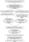
Figure 1
Figure 1
Schematic summarizing the overall study design. A stepwise approach was followed to select 6 genes for an association study in cases of MI and controls. The results of the association study prompted a study to determine the effect of the silencing of 2 genes on thrombus formation in D rerio. The protein encoded by the gene with the strongest effect on thrombus formation was selected for proteomics analysis in human platelets. PFC, Platelet Function Cohort; CAD, coronary artery disease; WTCCC, Wellcome Trust Case Control Consortium; GWAS, genome-wide association study; SNP, single-nucleotide polymorphism. Inclusion of the 6th gene (GTF2A2) was based on association of expression with all 4 measures of the platelet response and association.
Figure 2
Figure 2
Platelet functional response in the Platelet Function Cohort. Comparison of the platelet response to ADP and CRP-XL in the 37 selected subjects (○) with the 500 subjects in the whole PFC (•). Platelet activation was measured by flow cytometry, and data are shown as the standardized residuals of logit transformed data, after adjustment for confounding variables, as described by Jones et al. PA, P-selectin expression in response to ADP; PC, P-selectin expression in response to CRP-XL; FA, fibrinogen binding in response to ADP; FC, fibrinogen binding in response CRP-XL. Error bars show the median and interquartile ranges.
Figure 3
Figure 3
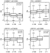
Genes showing association of transcript abundance in platelets with platelet response. (A) Sixty-three transcripts in platelets that show a correlation between microarray signal intensity (SI) and functional responses. Genes are grouped alphabetically and by cellular location of the encoded proteins. Platelet activation was measured as fibrinogen binding (F) and P-selectin expression (P) in response to ADP (A) or the collagen mimetic, CRP-XL (C). Correlation with functional responses for each transcript is indicated by the colored squares, with positive and negative correlations indicated by the diagonal lines. The 6 genes selected for genotyping in the cases of myocardial infarction (MI) and controls are highlighted in light blue and bold upper case. (B) Correlation between platelet transcript level and platelet function, in 37 subjects selected for the whole-genome expression study, for the 2 genes showing association with MI. SI was measured with Illumina probes GI_29789284-S and GI_4758689-S for COMMD7 and LRRFIP1, respectively. Platelet activation is shown as the standardized residuals of logit transformed flow-cytometric measurement of the percentage of platelets positive for specific markers of activation, as previously described. Graphs display the correlation with the highest significance, and boxes show Pearson correlation coefficients (r) and probability values (in parentheses) for all 4 functional parameters, with the highest significance shown in bold.
Figure 4
Figure 4
Box plots showing the effect of sequence variation in the COMMD7, LRRFIP1, and PFKL genes on the platelet response. Data are shown for the SNPs that showed the lowest P value in the PFC and the lowest P value in the replication MI/CAD cohort. The association between sequence variation and the platelet response is shown as the mean and interquartile range. Platelet activation was measured by flow cytometry, and data are shown as the standardized residuals of logit transformed data, after adjustment for confounding variables, as described by Jones et al. PA, P-selectin expression in response to ADP; FA, fibrinogen binding in response to ADP. Numbers (n) below graphs indicate the numbers of subjects homozygous for the major allele (1), heterozygous (2), or homozygous for the minor allele (3).
Figure 5
Figure 5
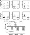
The effect of antisense knockdown of commd7, lrrfip1, and itga2b in D. rerio on thrombus kinetics. (A) Examples of results for the knockdown of commd7, lrrfip1, and itga2b (positive control) from 1 experiment showing the time to attachment of the first cell (TTA) in the top panels and the thrombus area (TA) in the bottom panels in control (•) and morpholino (MO)–injected (○) embryos. (B) Combined data for all experiments showing data as the percentage difference between control and the gene of interest (± confidence intervals) in either TTA (□) or TA (■). Injection of complementary MO resulted in a nonsignificant increase in TTA in the commd7 and itga2b morphants and a significant increase (P = .0035) in the lrrfip1 morphants. Knockdown of all 3 genes resulted in a reduction in TA of 30% (P = .027), 54% (P = .0004), and 55% (P = .0006) respectively. The effect of knockdown on TTA was estimated using Cox regression, and stratifying by experimental day and the effect on TA was estimated using a linear mixed-effects model as described in “Methods.”
Figure 6
Figure 6
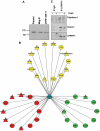
LRRFIP1 interactions in resting and activated human platelets imply a role in the regulation of the platelet cytoskeleton. (A) Western blotting with anti-LRRFIP1 identifies a 160-kDa protein in platelets and the megakaryocytic cell lines, Meg-01 and CHRF-288-11. (B) Tandem mass spectrometry analysis LRRFIP1 coimmunoprecipitation (co-IP) from resting and activated platelets identified novel and known protein-protein interactions (Table 2). Shown is a Cytoscape visualization of the LRRFIP1 protein-protein interaction network in platelets (Cytoscape Version 2.5). Node colors indicate interactions detected in resting only (green), activated only (red), or both resting and activated (yellow) platelets. LRRFIP1, which was detected in both resting and activated co-IPs; LRRFIP1 is shown centrally in blue. Node diameters indicate the number of peptide hits; a semiquantitative measure of protein abundance. (C) Western blotting of LRRFIP1 co-IPs with specific anti-Flightless 1 and anti-Drebrin antibodies confirmed these protein-protein interactions.

References

    1. Jones CI, Garner SF, Angenent W, et al. Mapping the platelet profile for functional genomic studies and demonstration of the effect size of the GP6 locus. J Thromb Haemost. 2007;5(8):1756–1765. - PubMed
    1. Panzer S, Hocker L, Koren D. Agonists-induced platelet activation varies considerably in healthy male individuals: studies by flow cytometry. Ann Hematol. 2006;85(2):121–125. - PubMed
    1. O'Donnell CJ, Larson MG, Feng D, et al. Genetic and environmental contributions to platelet aggregation: the Framingham Heart Study. Circulation. 2001;103(25):3051–3056. - PubMed
    1. Macaulay IC, Tijssen MR, Thijssen-Timmer DC, et al. Comparative gene expression profiling of in vitro differentiated megakaryocytes and erythroblasts identifies novel activatory and inhibitory platelet membrane proteins. Blood. 2007;109(8):3260–3269. - PMC - PubMed
    1. O'Connor MN, Salles II, Cvejic A, et al. Functional genomics in zebrafish permits rapid characterization of novel platelet membrane proteins. Blood. 2009;113(9):4754–4762. - PMC - PubMed

Publication types