. 2010 Nov 19;285(47):37005-15.
doi: 10.1074/jbc.M110.170076. Epub 2010 Sep 12.

Brain fatty acid-binding protein and omega-3/omega-6 fatty acids: mechanistic insight into malignant glioma cell migration

Affiliations

Brain fatty acid-binding protein and omega-3/omega-6 fatty acids: mechanistic insight into malignant glioma cell migration

Raja Mita et al. J Biol Chem. .

Abstract

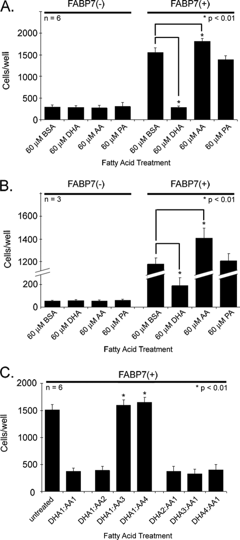
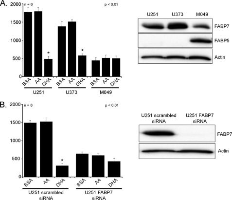
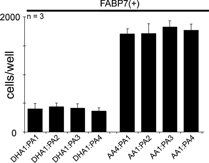
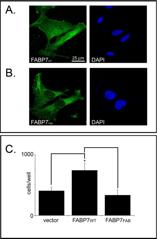
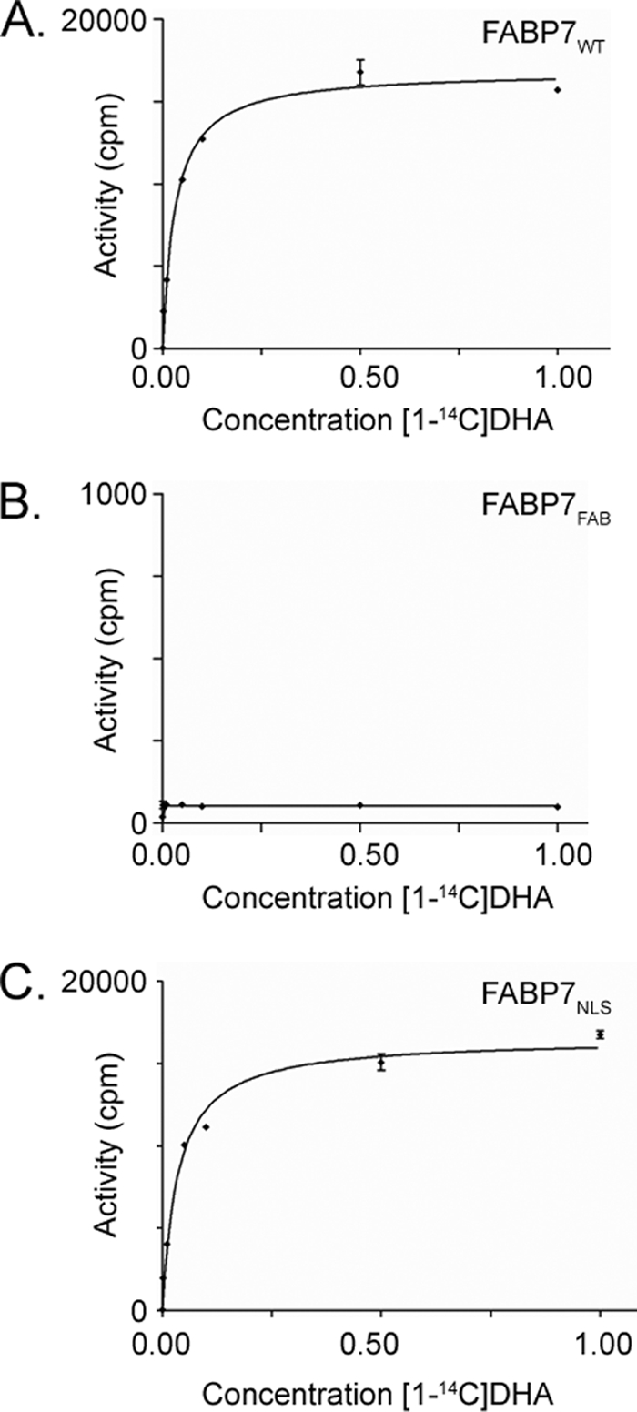
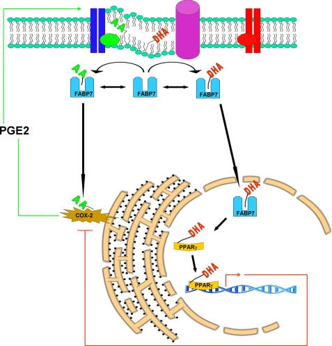
Malignant gliomas (MG) are highly infiltrative tumors that consistently recur despite aggressive treatment. Brain fatty acid-binding protein (FABP7), which binds docosahexaenoic acid (DHA) and arachidonic acid (AA), localizes to sites of tumor infiltration and is associated with a poor prognosis in MG. Manipulation of FABP7 expression in MG cell lines affects cell migration, suggesting a role for FABP7 in tumor infiltration and recurrence. Here, we show that DHA inhibits and AA stimulates migration in an FABP7-dependent manner in U87 MG cells. We demonstrate that DHA binds to and sequesters FABP7 to the nucleus, resulting in decreased cell migration. This anti-migratory effect is partially dependent on peroxisome proliferator-activated receptor γ, a DHA-activated transcription factor. Conversely, AA-bound FABP7 stimulates cell migration by activating cyclooxygenase-2 and reducing peroxisome proliferator-activated receptor γ levels. Our data provide mechanistic insight as to why FABP7 is associated with a poor prognosis in MG and suggest that relative levels of DHA and AA in the tumor environment can make a profound impact on tumor growth properties. We propose that FABP7 and its fatty acid ligands may be key therapeutic targets for controlling the dissemination of MG cells within the brain.


Figures

FIGURE 1.
FIGURE 1.
Cell migration of U87-FABP7(−) and U87-FABP7(+) cells in the presence of fatty acids. A, cells cultured in DMEM plus 10% FCS (growth medium) were treated with 60 μm BSA, 60 μm DHA, 60 μm AA, or 60 μm PA for 24 h. B, cells were serum-starved for 24 h and then treated with either BSA or the indicated fatty acids for 24 h. C, cells were cultured in growth medium and treated with different ratios of DHA and AA as follows: 1 = 30 μm; 2, = 60 μm; 3 = 90 μm; 4 = 120 μm. A–C, cells were trypsinized, and 20,000 cells (in DMEM) were plated in the upper chambers of Transwell inserts. The bottom chambers contained DMEM plus 10% FCS. After 6 h, the cells migrating through the porous membrane were fixed, stained, and counted using Metamorph imaging software version 7.7. Statistical significance was determined using the unpaired t test. Error bars represent standard deviation.
FIGURE 2.
FIGURE 2.
Cell migration of U251, U373, and M049 cells in the presence of fatty acids. A, FABP7(+) U251, U373, and M049 MG cells were cultured for 24 h in DMEM + 10% FCS containing 60 μm BSA, 60 μm DHA, or 60 μm AA. B, U251 cells transfected with control (scrambled) siRNA and FABP7 siRNA were cultured for 24 h in DMEM + 10% FCS containing 60 μm BSA, 60 μm DHA, or 60 μm AA. A and B, 20,000 cells (in DMEM) were plated in the upper chambers of Transwell inserts. The bottom chambers contained DMEM plus 10% FCS. After 6 h, the cells migrating through the porous membrane were fixed, stained, and counted using Metamorph imaging software version 7.7. Statistical significance was determined using the unpaired t test (*, p < 0.01). Error bars represent standard deviation. For Western blot analysis, proteins (25 μg/lane) were separated in a 13.5% SDS-polyacrylamide gel, transferred to nitrocellulose membranes, and immunostained with rabbit anti-FABP7 antibody, rabbit anti-FABP5 antibody, and mouse anti-actin antibody, followed by HRP-conjugated secondary antibodies.
FIGURE 3.
FIGURE 3.
Cell migration of U87-FABP7(+) cells treated with varying fatty acid ratios. U87-FABP7(+) cells were grown in DMEM plus 10% FCS and treated with varying fatty acid ratios for 24 h: 1 = 30 μm, 2 = 60 μm, 3 = 90 μm, 4 = 120 μm. Cell migration was measured using the Transwell assay. Twenty thousand cells/well were plated and incubated for 6 h, and the cells migrating through the porous membrane were fixed, stained, and counted using Metamorph imaging software. Statistical significance was determined using the unpaired t test. Error bars represent standard deviation.
FIGURE 4.
FIGURE 4.
PUFA-dependent subcellular localization of FABP7 in U87-FABP7(+) and U251MG cell lines. U87-FABP7(+) and U251MG cells expressing endogenous FABP7 were serum-starved for 24 h (A and D) and then were either treated with 60 μm DHA in the absence of FCS for an additional 24 h (B and E) or treated with 60 μm AA in the absence of FCS for 24 h (C and F). The subcellular localization of FABP7 in U87-FABP7(+) and U251MG was analyzed by immunofluorescence using anti-FABP7 antibody followed by Alexa 488-conjugated secondary antibody. The DNA was counterstained with DAPI.
FIGURE 5.
FIGURE 5.
Western blot analysis of FABP7 in U87-FABP7(+) stable transfectants treated with 60 μm BSA, AA, DHA, or PA. Whole cell lysates (25 μg/lane) were prepared from U87-FABP7(+) and electrophoresed in a 13.5% SDS-polyacrylamide gel. Proteins were transferred to nitrocellulose membranes and sequentially immunostained with rabbit anti-FABP7 antibody and mouse anti-actin antibody, followed by HRP-conjugated secondary antibodies. C, cytoplasmic; N, nuclear.
FIGURE 6.
FIGURE 6.
FABP7 levels in U87MG cells transfected with wild-type and mutant FABP7 expression constructs. U87MG cells were transfected with pcDNA3.1-FABP7WT, -FABP7NLS, and -FABP7FAB expression constructs. Cytoplasmic (C) and nuclear (N) fractions were prepared, and 25 μg of protein was loaded in each lane. Proteins were electrophoresed in a 13.5% SDS-polyacrylamide gel, transferred to nitrocellulose membranes, and immunostained with anti-FABP7 antibody. The signal was detected using ECL reagent (GE Healthcare.
FIGURE 7.
FIGURE 7.
Fatty acid binding is required for migration. Subcellular localization of FABP7 in U87MG cells transiently transfected with pcDNA3.1-FABP7WT (A) or pcDNA3.1-FABP7FAB (B) expression constructs. Transfection efficiency was ∼40%. Immunofluorescence was carried out 48 h after transfection using anti-FABP7 antibody followed by Alexa 488-conjugated secondary antibody. DNA was counterstained with DAPI. C, migration of U87MG cells transfected with empty vector, pcDNA3.1-FABP7WT, and pcDNA3.1-FABP7FAB was measured using the Transwell assay as described for Fig. 1. Statistical significance was determined using the unpaired t test (n = 3; *, p < 0.05). Error bars represent standard deviation.
FIGURE 8.
FIGURE 8.
Binding of DHA to FABP7WT, FABP7FAB, and FABP7NLS proteins. Reactions were carried out in the presence of different concentrations of [1-14C]DHA ranging from 0.001 to 5.0 μm (0.001, 0.01, 0.05, 0.1, 0.5, 1, and 5 μm) using 5 μg of recombinant FABP7WT (A), FABP7FAB (B), and FABP7NLS (C). Data points are the mean ± S.D. (n = 3).
FIGURE 9.
FIGURE 9.
FABP7-induced cell migration is independent of nuclear localization. A, U87MG cells were transfected with a pcDNA3.1-FABP7NLS expression construct. FABP7NLS localization was analyzed 48 h after transfection by immunofluorescence using anti-FABP7 antibody followed by Alexa 488-conjugated secondary antibody. DNA was counterstained with DAPI to illustrate the location of FABP7 relative to the nucleus. B, cell migration of U87-FABP7WT and U87-FABP7NLS cells treated with BSA, 60 μm AA, or 60 μm DHA was measured using the Transwell assay as described for Fig. 1. Statistical significance was determined using the unpaired t test (n = 6; *, p < 0.05). Error bars represent standard deviation.
FIGURE 10.
FIGURE 10.
COX-2 involvement in FABP7-induced cell migration. A, Western blot analysis of whole cell lysates (25 μg/lane) prepared from U87 cells stably transfected with FABP7 or empty vector. Lysates were electrophoresed in a 13.5% SDS-polyacrylamide gel, and the proteins were transferred to nitrocellulose membranes. Membranes were sequentially immunostained with goat anti-COX-2 antibody and mouse anti-actin antibody. B, ELISA was used to measure PGE2 levels in U87-FABP7(−) versus U87-FABP(+) cell populations. The data were obtained from two independent experiments measured in triplicate. No error bars are shown for U87-FABP7(+) as all the wells were saturated for PGE2. C, U87-FABP7(+) stable transfectants were treated 60 μm AA, 60 μm DHA, and/or 200 μm NS398, as indicated. Cell migration was measured using the Transwell assay as described previously. Statistical significance was determined using the unpaired t test (*, p < 0.01). Error bars represent standard deviation.
FIGURE 11.
FIGURE 11.
Role of PPARs in FABP7/FA-mediated cell migration. A, Western blot analysis of PPARα, PPARβ, and PPARγ in whole cell lysates (25 μg/lane) prepared from U87-FABP7(−) and U87-FABP7(+) cell populations. Lysates were electrophoresed in a 13.5% SDS-polyacrylamide gel. B, Western blot of U87-FABP7(+) cells transfected with scrambled siRNA (control), siRNAs targeting PPARβ, or PPARγ. C, 24 h after transfection, cells cultured in DMEM plus 10% FCS were treated with 60 μm BSA, 60 μm DHA, or 60 μm AA for an additional 24 h and subjected to the Transwell migration assay. The data were obtained from two independent experiments carried out in triplicate.
FIGURE 12.
FIGURE 12.
Model of cytoplasmic and nuclear roles for FABP7/fatty acids in cell migration. Cytosolic FABP7 can bind to either DHA or AA depending on the relative abundance and availability of these two fatty acids. Binding of FABP7 to AA induces cell migration through transfer of AA to the COX-2 pathway, whereby AA is metabolized into pro-migratory signaling molecules such as PGE2. Binding of FABP7 to DHA results in translocation of FABP7 to the nucleus where DHA is transferred to PPARγ, resulting in down-regulation of pro-migratory genes.

Similar articles

Cited by

References

    1. Lin C. L., Lieu A. S., Lee K. S., Yang Y. H., Kuo T. H., Hung M. H., Loh J. K., Yen C. P., Chang C. Z., Howng S. L., Hwang S. L. (2003) Surg. Neurol. 60, 402–406 - PubMed
    1. Mason W. P., Maestro R. D., Eisenstat D., Forsyth P., Fulton D., Laperrière N., Macdonald D., Perry J., Thiessen B. (2007) Curr. Oncol. 14, 110–117 - PMC - PubMed
    1. Godbout R., Bisgrove D. A., Shkolny D., Day R. S., 3rd (1998) Oncogene 16, 1955–1962 - PubMed
    1. Liang Y., Diehn M., Watson N., Bollen A. W., Aldape K. D., Nicholas M. K., Lamborn K. R., Berger M. S., Botstein D., Brown P. O., Israel M. A. (2005) Proc. Natl. Acad. Sci. U.S.A. 102, 5814–5819 - PMC - PubMed
    1. Tso C. L., Shintaku P., Chen J., Liu Q., Liu J., Chen Z., Yoshimoto K., Mischel P. S., Cloughesy T. F., Liau L. M., Nelson S. F. (2006) Mol. Cancer Res. 4, 607–619 - PubMed

Publication types

MeSH terms