. 2010 Oct 20;29(20):3520-30.
doi: 10.1038/emboj.2010.226. Epub 2010 Sep 10.

Crystal structure of Lon protease: molecular architecture of gated entry to a sequestered degradation chamber

Affiliations

Crystal structure of Lon protease: molecular architecture of gated entry to a sequestered degradation chamber

Sun-Shin Cha et al. EMBO J. .

Abstract

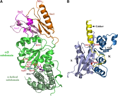
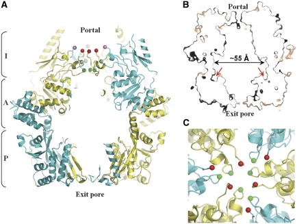
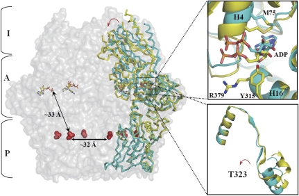
Lon proteases are distributed in all kingdoms of life and are required for survival of cells under stress. Lon is a tandem fusion of an AAA+ molecular chaperone and a protease with a serine-lysine catalytic dyad. We report the 2.0-Å resolution crystal structure of Thermococcus onnurineus NA1 Lon (TonLon). The structure is a three-tiered hexagonal cylinder with a large sequestered chamber accessible through an axial channel. Conserved loops extending from the AAA+ domain combine with an insertion domain containing the membrane anchor to form an apical domain that serves as a gate governing substrate access to an internal unfolding and degradation chamber. Alternating AAA+ domains are in tight- and weak-binding nucleotide states with different domain orientations and intersubunit contacts, reflecting intramolecular dynamics during ATP-driven protein unfolding and translocation. The bowl-shaped proteolytic chamber is contiguous with the chaperone chamber allowing internalized proteins direct access to the proteolytic sites without further gating restrictions.


Conflict of interest statement

The authors declare that they have no conflict of interest.

Figures

Figure 1
Figure 1
Overall structure of TonLon. (A) The hexameric structure of TonLon. The hexamer is shown with five subunits in surface representation and one monomer as a ribbon diagram. The molecule is oriented with the protease domain (P) (blue and lavender ribbons) at the bottom, the AAA+ domain (A) (bright and light green ribbons) in the middle, and the apical insertion domain (I) (orange and magenta ribbons) at the top. The orange ribbon depicts Insert 1, to which the membrane anchor (missing in our structure) is attached at the top. MA is the putative membrane-anchoring region. ADP is represented as a stick figure bound between the α/β (bright green) and α (light green) subdomains, and lavender balls indicate the positions of the protease catalytic residues. The other five monomers are in parallel alignment with T- and L-monomers in yellow and magenta tints, respectively. (B) Surface representation showing the top view of the hexamer, which has pseudo-six-fold symmetry. ADPs are shown in red. In subsequent figures, ‘T' (yellow) and ‘L' (cyan) represent T- and L-monomers, respectively.
Figure 2
Figure 2
Domain structure of TonLon. (A) Ribbon diagram showing the ATPase domain of a T-monomer. The α/β- and the α-helical subdomains are coloured in green and metallic green, respectively. Ins1, Ins2, and In3, which branch out of the α/β subdomain, are coloured in orange, magenta, and pink, respectively. MA is the putative membrane-anchoring region. Dots represent disordered regions in the final model. All residue numbers correspond to the sequence position in the full-length protein. The arginine finger (R311) is shown in stick. Important residues in the Walker-A (D245 and E246), Walker-B (K73), sensor-1 (N297), and sensor-2 (R379) motifs are shown in sticks and labelled. (B) Ribbon diagram showing the protease domain of T-monomers. The S-subdomain and the K subdomain, with catalytic K566, are labelled and are coloured in light blue and dark blue, respectively. A red ball indicates conserved glycine residue (G441 in TonLon and G596 in EcLon). The linker helix between the ATPase and protease domains is coloured yellow. Catalytic residues, S523 and K566, are shown as balls and labelled.
Figure 3
Figure 3
Clipped view of TonLon. (A) Ribbon drawing of a vertical section through the centre of the hexamer. Joining the AAA+ (I+A) and protease (P) domains forms a broad chamber with wide channels leading from the portal and to the exit pore. The portal opening is determined by two loops. F216 (red), in the centre of the aromatic-hydrophobic loop, and M275 (green), in the centre of the pre-sensor-1 β hairpin, are displaced downwards on the vertical axis in the T-monomers. (B) Surface rendering of a similar vertical slab. Red arrows point to the substrate-binding grooves in the protease layer, which are accessible to proteins from the chaperone portion of the chamber. (C) Top view of the hexamer in a ribbon drawing. Red and green spheres indicate Cα atoms of F216 and M275, respectively. These residues are closer to the vertical axis in the T-monomers.
Figure 4
Figure 4
Sequence alignment between α/β subdomains of TonLon and EcLon. The secondary structure assignments correspond to TonLon. A red arrow indicates the position of Ins1 (residues 85–189), which is shown in full in Supplementary Figure S1. Red and black open boxes frame Ins2 and Ins3, respectively. The Walker-A, Walker-B, and sensor-1 motifs are framed by blue, green, and yellow boxes, respectively. The arginine finger (R311) is shaded in pink. Asterisks indicate identical residues between TonLon and EcLon. The aromatic-hydrophobic loop motif of EcLon (Martin et al, 2008) is in bold letters.
Figure 5
Figure 5
Cα-tracing of superimposed T- and L-monomers. A T-monomer is superimposed onto an L-monomer in the hexamer, which is represented by a transparent surface. The protease domains were optimally aligned to emphasize the relative change in position of the AAA+ domain in the T-monomer, which is displaced down and in towards the molecular symmetry axis. The boxed region on the top right is the close-up view of the ATP-binding sites in the two monomers. The boxed region on the bottom right shows the pivot point near T323 about which the α/β subdomain rotates in the transition between the T and L states.
Figure 6
Figure 6
A close-up view of the TL interface. (A) At the interface between AAA+ domains, a modelled ATP is in stick representation and veiled by a transparent surface. A curved red arrow represents modelled rotation of the side chain of R311 upon ATP binding. Important residues in the Walker-B, sensor-1, and sensor-2 motifs are represented as sticks. Dotted lines point out interatomic distances <3 Å indicative of bonding. (B) The regions surrounding the linker helices of T- and L-monomers (yellow and cyan ribbons, respectively) are shown. Purple sticks represent residues from the adjacent L-monomer that come within 3 Å of the T-monomer linker. Residues within the protease domain that are displaced towards the T-monomer linker of the same subunit are shown as sticks. Linker residues whose side chains adopt different orientations in the two monomers are labelled green.

Similar articles

Cited by

References

    1. An YJ, Lee CR, Supangat S, Lee HS, Lee JH, Kang SG, Cha SS (2010) Crystallization and preliminary X-ray crystallographic analysis of Lon from Thermococcus onnurineus NA1. Acta Crystallogr Sect F Struct Biol Cryst Commun 66: 54–56 - PMC - PubMed
    1. Augustin S, Gerdes F, Lee S, Tsai FT, Langer T, Tatsuta T (2009) An intersubunit signaling network coordinates ATP hydrolysis by m-AAA proteases. Mol Cell 35: 574–585 - PMC - PubMed
    1. Baumeister W, Walz J, Zuhl F, Seemuller E (1998) The proteasome: paradigm of a self-compartmentalizing protease. Cell 92: 367–380 - PubMed
    1. Bieniossek C, Niederhauser B, Baumann UM (2009) The crystal structure of apo-FtsH reveals domain movements necessary for substrate unfolding and translocation. Proc Natl Acad Sci USA 106: 21579–21584 - PMC - PubMed
    1. Bieniossek C, Schalch T, Bumann M, Meister M, Meier R, Baumann U (2006) The molecular architecture of the metalloprotease FtsH. Proc Natl Acad Sci USA 103: 3066–3071 - PMC - PubMed

Publication types

Associated data

LinkOut - more resources