Comparative Study

Candidate exome capture identifies mutation of SDCCAG8 as the cause of a retinal-renal ciliopathy

Edgar A Otto et al. Nat Genet. 2010 Oct.

Abstract

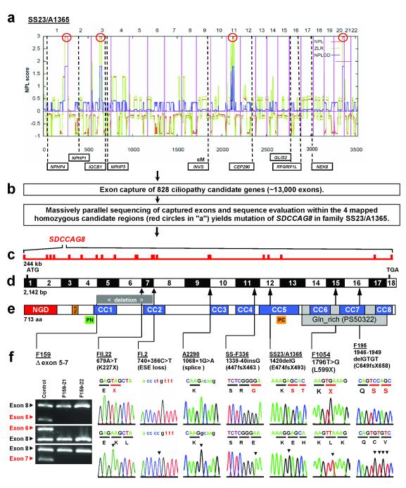
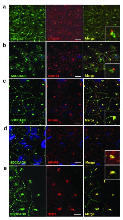
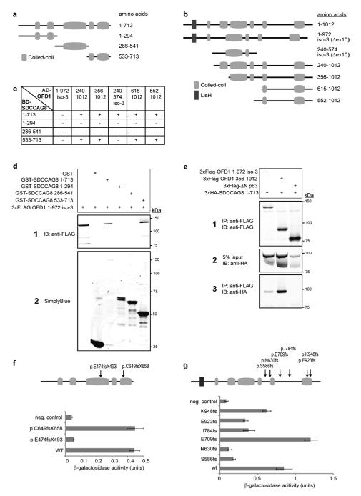
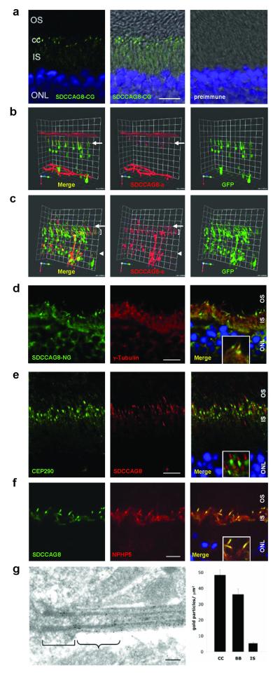
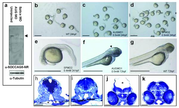
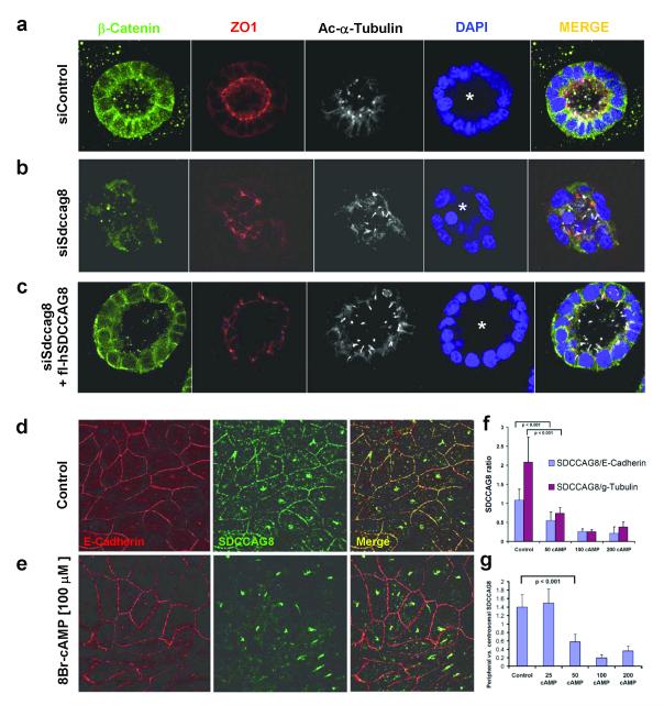
Nephronophthisis-related ciliopathies (NPHP-RC) are recessive disorders that feature dysplasia or degeneration occurring preferentially in the kidney, retina and cerebellum. Here we combined homozygosity mapping with candidate gene analysis by performing 'ciliopathy candidate exome capture' followed by massively parallel sequencing. We identified 12 different truncating mutations of SDCCAG8 (serologically defined colon cancer antigen 8, also known as CCCAP) in 10 families affected by NPHP-RC. We show that SDCCAG8 is localized at both centrioles and interacts directly with OFD1 (oral-facial-digital syndrome 1), which is associated with NPHP-RC. Depletion of sdccag8 causes kidney cysts and a body axis defect in zebrafish and induces cell polarity defects in three-dimensional renal cell cultures. This work identifies loss of SDCCAG8 function as a cause of a retinal-renal ciliopathy and validates exome capture analysis for broadly heterogeneous single-gene disorders.


Figures

Figure 1
Figure 1. Homozygosity mapping, exon capture, and massively parallel sequencing identifies SDCCAG8 mutations as causing nephronophthisis with retinal degeneration
(a) Non-parametric LOD (NPL) scores across the human genome in 2 sibs with nephronophthisis and retinal degeneration of consanguineous family SS23/A1365. X-axis gives Affymetrix 250k StyI array SNP positions on human chromosomes concatenated from p-ter (left) to q-ter (right). Genetic distance is given in cM. Four maximum NPL peaks (red circles) indicate candidate regions of homozygosity by descent. (b) Exon capture of 828 ciliopathy candidate genes with consecutive massively parallel sequencing and sequence evaluation within the 4 mapped homozygous candidate regions (red circles in “a”) yields mutation of SDCCAG8 in SS23/A1365. (c) The SDCCAG8 gene extends over 244 kb and contains 18 exons (vertical hatches). (d) Exon structure of human SDCCAG8 cDNA. Positions of start codon (ATG) and of stop codon (TGA) are indicated. For mutations detected (see f ) arrows indicate positions relative to exons and protein domains (see e). (e) Domain structure of the SDCCAG8 protein. N-terminal globular domain (NGD), nuclear localization (NLS) domain, coiled-coil domains (CC), and glutamine-rich region (Gln_rich). PN and PC denote peptides used for antibody generation. (f) Eight homozygous SDCCAG8 mutations detected in 8 families with nephronophthisis and retinal degeneration. Family number, mutation and predicted translational changes are indicated (see Table 1). A homozygous deletion covering exons 5-7 is demonstrated by agarose gel electrophoresis. Sequence traces are shown for mutations above normal controls. Mutated nucleotides are indicated by arrow heads in traces of normal controls.
Figure 2
Figure 2. Indirect immunofluorescence detects SDCCAG8 at centrosomes together with other proteins mutated in NPHP-RC
(a) Centrosomal SDCCAG8 is at centrosomes but slightly set apart from the signal of γ-tubulin, which marks the centrioles and from CEP164 (b), which marks distal centrosomal appendages. (c) In contrast, there is tight colocalization with ninein, a marker of centrosomal appendages. There is also colocalization at centrosomes with NPHP5 (d) and OFD1 (e) previously found to be mutated in NPHP-RC,. Size bars represent 5 μm. Insets show enlargement of representative results at 5-fold higher magnification. Nuclei are stained with DAPI. Antibody α-SDCCAG8-CG was used to detect SDCCAG8.
Figure 3
Figure 3. SDCCAG8 Interaction with OFD1
(a, b) Schematic of SDCCAG8 and OFD1 clones and protein domains. Full-length SDCCAG8 (Q86SQ7) and three cloned fragments are shown (a) as well as OFD1 fragments identified by yeast two-hybrid screening and isoforms O75665-1 and O75665-3. (c) Yeast two-hybrid interaction assays confirmed binding of full-length SDCCAG8 and C-terminal fragment (533-713) to all cloned OFD1 fragments, except isoform-3. HIS3 and ADE2 reporter genes activation is indicated. (d) Both GST-SDCCAG8 full-length and the GST-SDCCAG8 C-terminal fragment efficiently pulled down 3xFLAG-tagged OFD1 isoform-3, contrary to GST alone. (e) 3xHA-tagged full-length SDDCAG8 coimmunoprecipitated with 3xFLAG-tagged OFD1 isoform 3, and fragment 356-1012, contrary to unrelated 3xFLAG-tagged ΔN-p63. Panel 2 shows 5% of cell lysate input. (f) Interaction of wild-type (WT) and SDCCAG8 mutants with ODF1 isoform-1 in beta-galactosidase assay. Constructs encoding SDCCAG8 as GAL4-BD fusion protein, WT or indicated mutants, were cotransformed with pAD-OFD1 isoform-3 constructs in yeast. Cotransformation of pBD-SDCCAG8 and pAD-MUT vector served as negative control. Remaining LacZ reporter gene activity, corrected for background activity, is indicated. (g) Interaction of WT isoform-3 and OFD1 mutants with SDCCAG8 in yeast two-hybrid assay. Constructs encoding WT SDCCAG8 as GAL4-BD fusion protein were cotransformed in PJ694a with pAD-OFD1 isoform-3 constructs, WT or indicated mutations. Interaction of SDCCAG8 with OFD1 mutant p.E709fs and WT was detected as described above. Beta-galactosidase assays revealed decreased LacZ reporter gene activity for OFD1 all mutations except E709fs, indicating significantly reduced interaction with SDCCAG8. Error bars represent standard error of the mean in f and g.
Figure 4
Figure 4. SDCCAG8 is located in mouse photoreceptor basal bodies and connecting cilia transition zone
(a) Immunofluorescence using the α-SDCCAG8-CG antibody demonstrates strong SDCCAG8 expression in transition zone of connecting cilia (cc) and weak expression in inner segments (IS) of mouse photoreceptors. (b-c) Upon in vivo electroporation into rat retinae, human full-length SDCCAG8 isoform-a (red) (b) localizes to transition zone of photoreceptor cells (level of arrow), whereas the short N-terminal isoform-e (red) (c) exhibits expression in transition zone (level of arrow), inner segments (level of bracket) and cytoplasm (level of arrow head). Photoreceptor cytoplasms are counterstained by GFP overexpression (green). (d) In mouse photoreceptors SDCCAG8 is located in transition zone of connecting cilia, distal to basal body marker γ-tubulin in a contiguous but non-overlapping location. (e) SDCCAG8 is located in the transition zone, distal to, and clearly set apart from the pericentriolar marker CEP290. (f) SDCCAG8 tightly colocalizes with NPHP5. Scale bars are 10 μm. Insets in d-f show enlargement of representative results at 3-fold higher magnification. (g) Upon ultrathin sectioning of mouse retina SDCCAG8 expression is particularly prominent at distal basal body (bracket) and the transition zone (winged bracket) connecting cilium of mouse photoreceptors (antibody α-SDCCAG8-PR). The bar graph represents the density of immunogold labeling over the photoreceptor transition zone (TZ), basal bodies (BB) and inner segments (IS) after subtracting background labeling. Scale bar = 100 nm.
Figure 5
Figure 5. sdccag8 knockdown results in multiple developmental defects
(a) Injection of both sdccag8-targeting MOs abrogates sdccag8 protein from MO injected embryo morphant lysates. sdccag8 protein is detected by the α-SDCCAG8-NR antibody as a single band in uninjected embryos, compatible with the sdccag8 full-length product at ~83 kDa (arrow head). Anti-α-tubulin antibody was used to demonstrate equal loading. (b-d) In comparison to embryos injected at the 2-cell stage with standard negative control MO (b) embryos injected with AUG-targeting MO (c) or splice site targeting MO (d,e) exhibit a dose-dependent phenotype of body axis curvature and shortened and broadened tails at 24 hpf. (f-k) Knockdown of sdccag8 in zebrafish embryos at 72 hpf caused pronephric cysts as evidenced by a rounded structure (arrow in f) and hollow spaces (asterisk in h) compared to control morpholino injected control (g,j) , which shows slender pronephric tubular lumina (arrows in i). It also caused hydrocephalus (asterisk in j) compared to control morpholino injected control (g,k). Scale bars represent 1 mm in b-g and 100 μm in h-k.
Figure 6
Figure 6. siRNA knockdown of siSdccag8 perturbs lumen formation of renal epithelial cells in 3D spheroid culture
(a) IMCD3 murine renal epithelial cells, when grown in 3D matrigel culture for 3 days, polarize as demonstrated by staining for β-catenin (green) and tight junction marker ZO1 (red). They ciliate apically as shown by acetylated α-tubulin (white) and form a spheroid containing a central lumen (asterisk). Nuclei are stained blue with DAPI. (b) Upon siSdccag8 knockdown cells develop spheroids that have architectural defects characterized by disturbed localization of β-catenin (green) away from the basolateral membrane, fewer tight junctions (red), and an irregular lumen (asterisk). Abnormal lumina were seen in 19.5% of siRNA controls versus 75.8% in siSdccag8 knockdown (p=0.0055). (c) When siSdccag8 knockdown was performed in mIMCD3 cells that were stably transfected with human full-length SDCCAG8, the knockdown phenotype was fully rescued leading to no significant reduction in irregular lumina (16.7%) compared to negative control siRNA (19.5%). (d-g) SDCCAG8 abandons cell-cell junctions in response to increased intracellular cAMP. (d) In the renal epithelial cell line MCDK-II SDCCAG8 stained with antibody α-SDCCAG8-CG (green) is located at centrosomes and cell-cell junctions, which are marked with an α-E-cadherin antibody (red). (e) Upon treatment with 8-Br-cAMP [100 μM], there is dose-dependent loss of SDCCAG8 signal from cell-cell junctions relative to E-cadherin (f-g). When 100 linear segments of the pentagonal cell-cell junctions of non-neighboring cells were evaluated for the ratio of relative fluorescence signals for SDCCAG8 versus E-cadherin there was a redistribution depending on cAMP concentration [μM] away from cell junction staining for SDCCAG8 relative to peripheral E-cadherin, centrosomal γ-tubulin (f) and relative to centrosomal SDCCAG8 (g) (see also Supplementary Figure 6 ). Error bars represent standard error of the mean.

Comment in

References

    1. Hildebrandt F, et al. A novel gene encoding an SH3 domain protein is mutated in nephronophthisis type 1. Nat Genet. 1997;17:149–153. - PubMed
    1. Otto EA, et al. Mutations in INVS encoding inversin cause nephronophthisis type 2, linking renal cystic disease to the function of primary cilia and left-right axis determination. Nat Genet. 2003;34:413–420. - PMC - PubMed
    1. Olbrich H, et al. Mutations in a novel gene, NPHP3, cause adolescent nephronophthisis, tapeto-retinal degeneration and hepatic fibrosis. Nat Genet. 2003;34:455–459. - PubMed
    1. Otto E, et al. A gene mutated in nephronophthisis and retinitis pigmentosa encodes a novel protein, nephroretinin, conserved in evolution. Am J Hum Genet. 2002;71:1167–1171. - PMC - PubMed
    1. Mollet G, et al. The gene mutated in juvenile nephronophthisis type 4 encodes a novel protein that interacts with nephrocystin. Nat Genet. 2002;32:300–305. - PubMed

Publication types

MeSH terms

Associated data