. 2010 Oct;13(10):1233-9.
doi: 10.1038/nn.2637. Epub 2010 Sep 12.

Clarke's column neurons as the focus of a corticospinal corollary circuit

Affiliations

Clarke's column neurons as the focus of a corticospinal corollary circuit

Adam W Hantman et al. Nat Neurosci. 2010 Oct.

Abstract

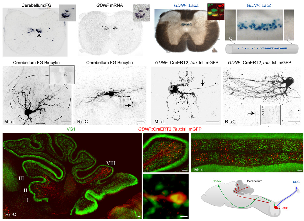
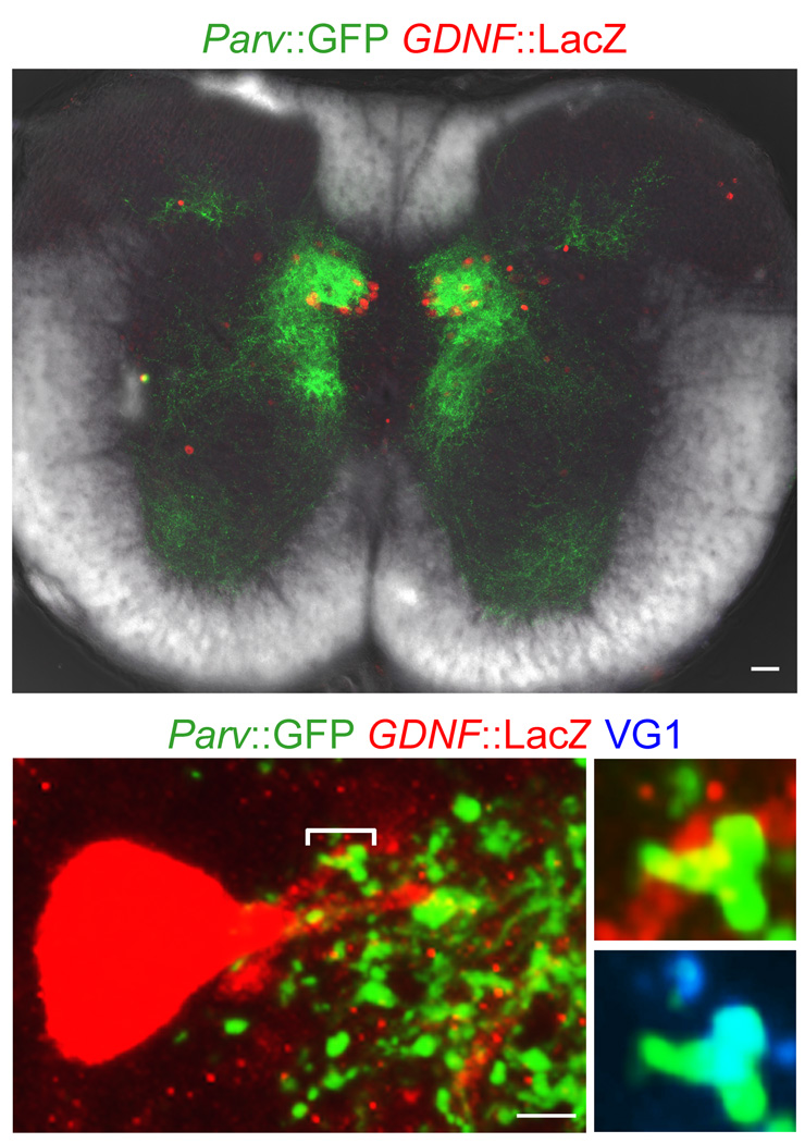
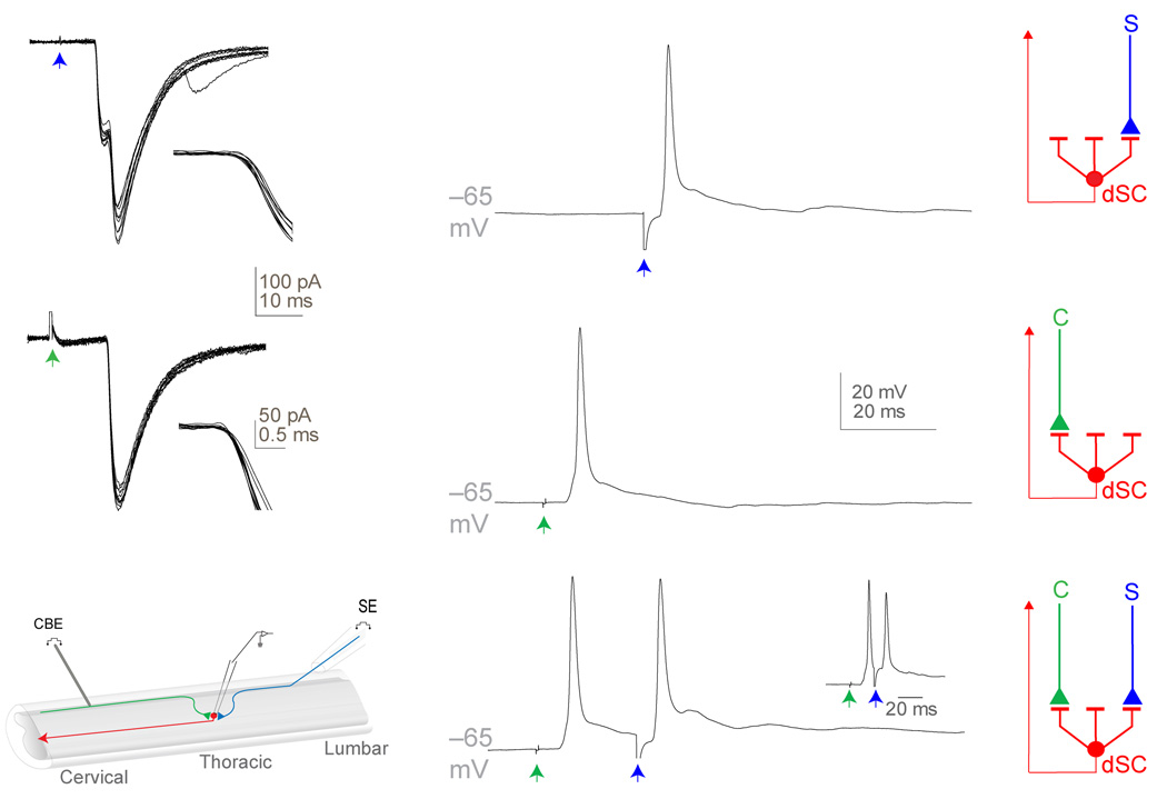
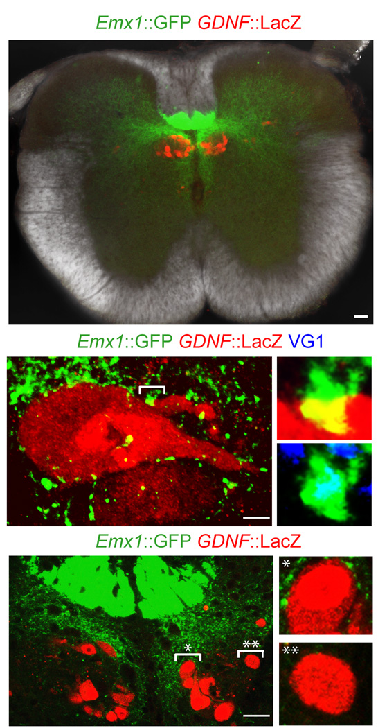
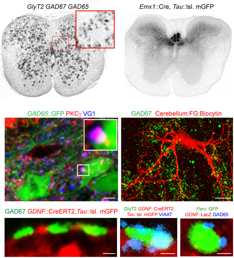
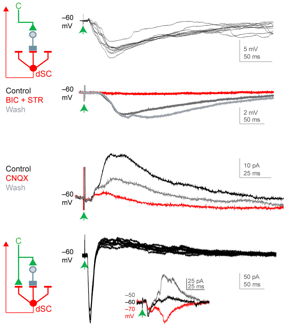
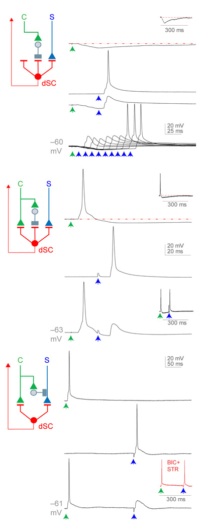
Proprioceptive sensory signals inform the CNS of the consequences of motor acts, but effective motor planning involves internal neural systems capable of anticipating actual sensory feedback. Just where and how predictive systems exert their influence remains poorly understood. We explored the possibility that spinocerebellar neurons that convey proprioceptive sensory information also integrate information from cortical command systems. Analysis of the circuitry and physiology of identified dorsal spinocerebellar tract neurons in mouse spinal cord revealed distinct populations of Clarke's column neurons that received direct excitatory and/or indirect inhibitory inputs from descending corticospinal axons. The convergence of these descending inhibitory and excitatory inputs to Clarke's column neurons established local spinal circuits with the capacity to mark or modulate incoming proprioceptive input. Together, our genetic, anatomical and physiological results indicate that Clarke's column spinocerebellar neurons nucleate local spinal corollary circuits that are relevant to motor planning and evaluation.


Figures

Figure 1
Figure 1. Anatomic and genetic characterization of post-natal mouse dSC neurons
(a) Location of thoracic spinal neurons retrogradely-labeled from cerebellum with fluorogold (FG). Clarke’s column marked by arrowhead; inset, high-magnification of FG in Clarke’s column. (b) GDNF mRNA expression in thoracic spinal cord (inset, GDNF expression in Clarke’s column of a inset). A small group of retrogradely-labeled, GDNF-expressing neurons are also found in the deep dorsal horn. (c) LacZ activity in GDNF::LacZ spinal cord (inset, co-labeling of LacZ and retrograde fluorogold label from the cerebellum in Clarke’s column). (d) Upper, GDNF neurons along the rostral-caudal axis (C, cervical; T, thoracic; L, caudal lumbar). Lower, summary of dSC distribution. High density in caudal thoracic levels; low density in rostral thoracic; and absent in cervical, caudal lumbar, and sacral (not shown) levels. (e) Morphology of a biocytin-filled, fluorogold+ dSC neuron in a transverse section (inset, low-magnification image; scale bar, 25µm) and (f) in a sagittal section (arrow marks axon; scale bar, 50µm). (g), Morphology of GDNF::CreERT2, Tau::lsl. mGFP+ dSC neurons (arrow marks axon) in a transverse section and (h) in a sagittal section (arrow marks axon; inset, horizontal spinal section of dorsal lateral funiculus). Scale bars g,h; 25µm. i, GDNF::CreERT2, Tau::lsl. mGFP+ dSC axons in a sagittal section of cerebellum. (j) Magnified image of GDNF::CreERT2, Tau::lsl. mGFP+ dSC axons of lobule VIII of i. (k) GDNF::CreERT2, Tau::lsl. mGFP+ dSC axons in a coronal section of cerebellum. Scale bars i–k, 50µm. (l) VG1 expression in GDNF::CreERT2, Tau::lsl. mGFP+ dSC axons. Scale bar, 1µm. (m) Summary of dSC anatomy (DRG, dorsal root ganglia).
Figure 2
Figure 2. Anatomy of proprioceptive inputs to dSC neurons
(a) Low-magnification image of thoracic spinal cord including Parv::GFP+ expressing proprioceptor terminals and GDNF::LacZ+ dSC neurons. Scale bar, 50µm. (b) Parv::GFP+ expressing proprioceptor terminals on an individual GDNF::LacZ+ dSC neuron (right-top, high-magnification image of bracketed area; right-bottom, Parv and VG1 expression of bracketed area). Scale bar, 5µm.
Figure 3
Figure 3. Physiology of proprioceptive and cortical inputs to dSC neurons
(a) 10 dorsal root (blue arrow)-evoked EPSCs in a dSC neuron (inset, expanded time scale around response onset). (b) Single dorsal root-evoked EPSP in a dSC neuron, dorsal root-evoked action potentials were detected in 26/31 neurons. Schematic of sensory (S, blue) input to dSC neurons. (c) 10 dorsal column (green arrow)-evoked EPSCs in a dSC neuron (inset, expanded time scale around response onset). (d) Single dorsal column-evoked EPSP in a dSC neuron, dorsal column-evoked action potentials were generated in 14/20 neurons. Schematic of cortical (C, green) input to dSC neurons. (e) Hemisected spinal cord preparation. Lumbar (L4–L6) dorsal roots (blue) were stimulated with a suction electrode (SE) and cervical dorsal column (green) was stimulated with a concentric bipolar electrode (CBE). (f) EPSPs of dSC neurons recorded after dorsal root and dorsal column stimulation (inset, reduced interstimulus interval between dorsal root and dorsal column stimulation). Schematic of convergence of cortical and sensory inputs on dSC neurons.
Figure 4
Figure 4. Anatomy of cortical inputs to dSC neurons
(a) Low-magnification image of thoracic spinal cord including Emx1::GFP+ expressing corticospinal terminals and GDNF::LacZ+ dSC neurons. Scale bar, 50µm. (b) Emx1::GFP+ corticospinal terminals on an individual GDNF::LacZ+ dSC neuron (right-top, high-magnification image of bracketed area; right-bottom, GFP and VG1 expression of bracketed area). Scale bar, 5µm. (c) Organization of Emx1::GFP+ corticospinal terminals with respect to location of GDNF::LacZ+ dSC neurons (right-top, magnified image of * bracketed area; right-bottom, magnified image of ** bracketed area). Scale bar, 25µm.
Figure 5
Figure 5. Anatomy of cortically-evoked inhibition of dSC neurons
(a) In situ hybridization of GlyT2, GAD67, and GAD65 probes in thoracic spinal cord (inset, magnified image of boxed area). (b) Distribution of Emx1::Cre, Tau::lsl. mGFP+ corticospinal terminals in thoracic spinal cord. (c) Apposition of PKC-γ+, VG1+ corticospinal terminals and GAD65::GFP+ neurons (inset, magnified image of boxed area). Scale bar, 5µm. (d) GAD67+ inhibitory inputs on a biocytin-filled dSC neuron. Scale bar, 50µm. (c) GABAergic (GAD67+) and (f) glycinergic (GlyT2+ and VIAAT+) inhibitory inputs on GDNF::CreERT2, Tau::lsl. mGFP+ dSC neurons. (g) GAD65+ inhibitory terminals on Parv::GFP+ proprioceptive terminals in Clarke’s column. Scale bars e–g, 1µm.
Figure 6
Figure 6. Physiology of cortically-evoked inhibition of dSC neurons
(a) Left, schematic of cortical excitation (green) of an inhibitory input (gray) to dSC neurons. Right, 10 dorsal column-evoked IPSPs in a dSC neuron. (b) Pharmacological assessment of bicuculline (BIC, GABAA-receptor antagonist, 8 µM) and strychnine (STR, glycine-receptor antagonist, 10 µM) action on dorsal column-evoked IPSPs in a dSC neuron (each trace is average of 10 trials). (c) Pharmacological assessment of CNQX (AMPA-receptor antagonist, 10 µM) action on dorsal column-evoked IPSCs of a dSC neuron (each trace is average of 10 trials). (d) Left, schematic of cortical excitation (green) and inhibition (gray) of a dSC neuron. Right, 10 dorsal column-evoked EPSCs and IPSCs in a dSC neuron (inset, reversal potential of inhibitory component).
Figure 7
Figure 7. Cortical inhibition of sensory-evoked responses in dSC neurons
Three modes of dorsal column inhibition of dorsal root responses in dSC neurons. (a) Top, dorsal column-evoked IPSP in a dSC neuron (inset, longer time scale to show duration of inhibition); second, dorsal root-evoked EPSPs in a dSC neuron; third, dorsal root-evoked EPSP preceded by dorsal column-evoked IPSP; bottom, dorsal root input preceded by dorsal column input at different time intervals. (b) Top, dorsal column-evoked EPSP and IPSP in a dSC neuron (inset, longer time scale to show duration of inhibition); middle, dorsal root-evoked EPSP in a dSC neuron; bottom, dorsal root-evoked EPSP preceded by dorsal column-evoked EPSP and IPSP (inset, longer inter-stimulus interval between dorsal root and dorsal column stimulation). (c) Top, dorsal column-evoked EPSP in a dSC neuron; middle, dorsal root-evoked EPSP in a dSC neuron; bottom, dorsal root-evoked EPSP preceded by dorsal column-evoked EPSP (inset, dorsal root input preceded by dorsal column input in the presence of BIC and STR). All traces represent single trials. No IPSPs were detected in this cell, even at depolarized holding potentials. These findings are suggestive of a presynaptic inhibitory mechanism, which could also occur in neurons exhibiting cortically- evoked postsynaptic inhibition.

Comment in

References

    1. Eccles JC, Oscarsson O, Willis WD. Synaptic action of group I and II afferent fibres of muscle on the cells of the dorsal spinocerebellar tract. J Physiol. 1961;158:517–543. - PMC - PubMed
    1. Lundberg A. Ascending Spinal Hindlimb Pathways in the Cat. Prog Brain Res. 1964;12:135–163. - PubMed
    1. Oscarsson O. Functional Organization of the Spino- and Cuneocerebellar Tracts. Physiol Rev. 1965;45:495–522. - PubMed
    1. Bosco G, Poppele RE. Proprioception from a spinocerebellar perspective. Physiol Rev. 2001;81:539–568. - PubMed
    1. Matsushita M, Hosoya Y. Cells of origin of the spinocerebellar tract in the rat, studied with the method of retrograde transport of horseradish peroxidase. Brain Res. 1979;173:185–200. - PubMed

Publication types

MeSH terms

Substances