. 2010 Jun 18:4:123-56.
doi: 10.2174/1874357901004030123.

HSV Recombinant Vectors for Gene Therapy

Affiliations

HSV Recombinant Vectors for Gene Therapy

Roberto Manservigi et al. Open Virol J. .

Abstract

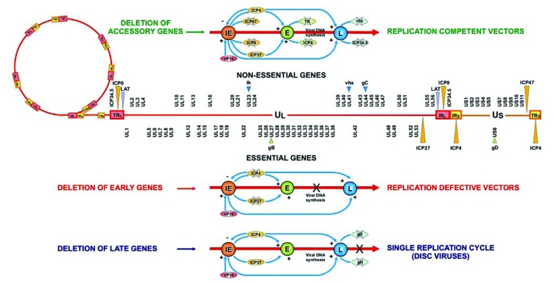
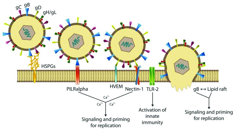
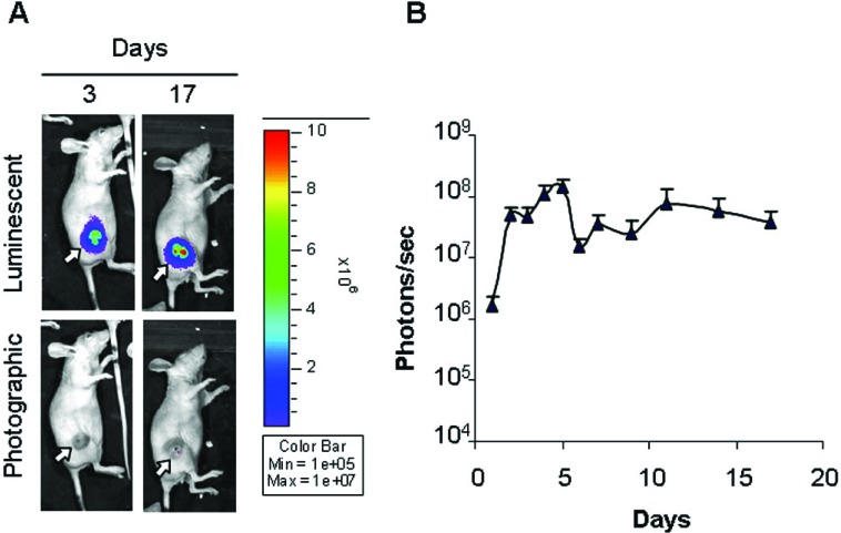
The very deep knowledge acquired on the genetics and molecular biology of herpes simplex virus (HSV), has allowed the development of potential replication-competent and replication-defective vectors for several applications in human healthcare. These include delivery and expression of human genes to cells of the nervous systems, selective destruction of cancer cells, prophylaxis against infection with HSV or other infectious diseases, and targeted infection to specific tissues or organs. Replication-defective recombinant vectors are non-toxic gene transfer tools that preserve most of the neurotropic features of wild type HSV-1, particularly the ability to express genes after having established latent infections, and are thus proficient candidates for therapeutic gene transfer settings in neurons. A replication-defective HSV vector for the treatment of pain has recently entered in phase 1 clinical trial. Replication-competent (oncolytic) vectors are becoming a suitable and powerful tool to eradicate brain tumours due to their ability to replicate and spread only within the tumour mass, and have reached phase II/III clinical trials in some cases. The progress in understanding the host immune response induced by the vector is also improving the use of HSV as a vaccine vector against both HSV infection and other pathogens. This review briefly summarizes the obstacle encountered in the delivery of HSV vectors and examines the various strategies developed or proposed to overcome such challenges.

Keywords: HSV; cancer; gene therapy; neurodegenerative disorders; oncolytic vectors; targeting; vaccines.; viral vectors.


Figures

Fig. (1)
Fig. (1)
Map of HSV-1 genome and schematic representation of replication-competent and defective vectors. A schematic representation of the position of the genes encoding proteins involved in virus replication, regulation, in virus formation and assembly, and in virion structural proteins is depicted. Genes that are modified or deleted to achieve viral attenuation are indicated.
Fig. (2)
Fig. (2)
Mechanism of HSV-1 entry into the host cell. The initial contact of the virus with the cell is the binding to the heparan sulfate (HS) proteoglycans on the cell surface, mediated by gC and gB, with consequent binding of gB to the PILRalpha receptor. Subsequently, gD binds to one of its cellular receptors, including HVEM, a member of the TNF-receptor family; nectin-1 or 2, two related members of the immunoglobulin superfamily; or sites generated in HS by the action of specific 3-O sulfotransferases. This last binding triggers the fusion between the cell membrane and the viral envelope, which requires the action of gB, gD and gH-gL, with subsequent release of the viral nucleocapsid and tegument into the cytoplasm. Gene therapy strategies aimed to target viral infection to particular cells can be obtained by modifying the first steps of the virus life cycle, that is, adsorption and penetration. The three main glycoproteins involved in these two phases are gB, gC and gD and their ORF backbone has been engineered to redirect infection to the target cell by deleting regions that affect binding to the main HSV receptors and/or inserting ligands that favour interaction with the new receptors. Envelope-HSV glycoproteins may also interact with TLRs on the cell surface, triggering signals that stimulate innate immunity.
Fig. (3)
Fig. (3)
Monitoring of virus transduction in a model of subcutaneous hepatocellular carcinoma (HCC) [367]. To follow HSV-1 C-gal-Luc strain replication in the tumour mass the virus was inoculated into the experimental tumour, developed on the right flank of athymic mice, and localization and intensity of luciferase expression was monitored by in vivo bioluminescence imaging. (A) Overlay luminescent/photographic and photographic images of a representative animal at 3 and 17 days post-infection. The arrows indicate the location of tumours. Intensity of light emission is represented by an artificial colour code normalized to allow comparison of different acquisitions. The maximum (red) and minimum (blue) correspond to 107 and 105 photons/s, respectively. (B) Quantification of luciferase activity over time. The average light emission in photons/s (n=7) is reported.

References

    1. Roizman B, Knipe DM, Whitley RJ. In: Fields Virology. 5th. Knipe DM, Howley PM, editors. Philadelphia, PA, Lippincot: Williams and Wilkins, a Wolters Kluwer Business; 2007. pp. 2501–601.
    1. Nicola AV, Straus SE. Cellular and viral requirements for rapid endocytic entry of herpes simplex virus. J Virol. 2004;78:7508–17. - PMC - PubMed
    1. Satoh T, Arii J, Suenaga T, et al. PILRalpha is a herpes simplex virus-1 entry coreceptor that associates with glycoprotein B. Cell. 2008;132:935–44. - PMC - PubMed
    1. Subramanian RP, Geraghty RJ. Herpes simplex virus type 1 mediates fusion through a hemifusion intermediate by sequential activity of glycoproteins D, H, L, and B. Proc Natl Acad Sci USA. 2007;104:2903–8. - PMC - PubMed
    1. Jardetzky TS, Lamb RA. Virology: a class act. Nature. 2004;427:307–8. - PubMed

LinkOut - more resources