. 2010 Nov 7;10(21):2911-6.
doi: 10.1039/c0lc00094a. Epub 2010 Sep 8.

Kinetics of NF-κB nucleocytoplasmic transport probed by single-cell screening without imaging

Affiliations

Kinetics of NF-κB nucleocytoplasmic transport probed by single-cell screening without imaging

Jun Wang et al. Lab Chip. .

Abstract

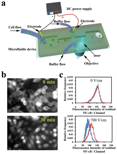
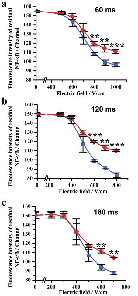
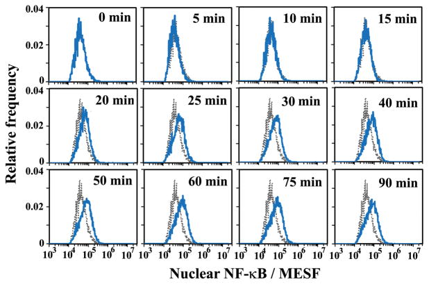
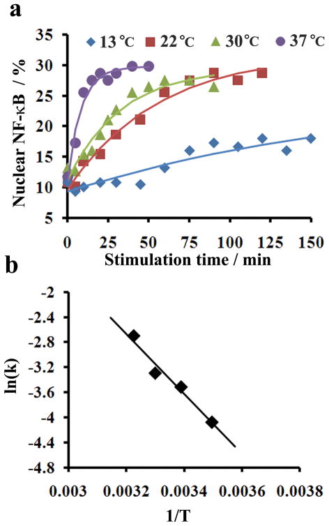
Transport of protein and RNA cargoes between the nucleus and cytoplasm (nucleocytoplasmic transport) is vital for a variety of cellular functions. The studies of kinetics involved in such processes have been hindered by the lack of quantitative tools for measurement of the nuclear and cytosolic fractions of an intracellular protein at the single cell level for a cell population. In this report, we describe using a novel method, microfluidic electroporative flow cytometry, to study kinetics of nucleocytoplasmic transport of an important transcription factor NF-κB. With data collected from single cells, we quantitatively characterize the population-averaged kinetic parameters such as the rate constants and apparent activation barrier for NF-κB transport. Our data demonstrate that NF-κB nucleocytoplasmic transport fits first-order kinetics very well and is a fairly reversible process governed by equilibrium thermodynamics.


Figures

Figure 1
Figure 1
(a) Schematic of the microfluidic electroporative flow cytometry device. The microfluidic device is mounted on a microscope that allows incident laser from a 40X objective to focus in the channel. A DC power supply generates a constant high electric field in the narrow sections to electroporate cells and release intracellular proteins. The width of the wide sections of the horizontal channel is 400 μm and that of the narrow section is 40 μm. The channel depth is 25 μm. (b) Fluorescence images of CHO cells expressing GFP labeled NF-κB. The cells were stimulated by 20 ng/ml IL-1 β at 37 °C at time 0. (c) Comparison of histograms of residual NF-κB between stimulated cells (red) and cells without stimulation (blue) under electric fields of 0 and 700 V/cm. The electroporation duration was 120 ms. The stimulation was conducted at 37 °C for 30 min.
Figure 2
Figure 2
The mean fluorescence intensity of a cell population generated by residual NF-κB after flow-through electroporation under different field intensities. Cell populations stimulated by IL-1β (red) and the ones without stimulation (blue) are compared when the field duration is (a) 60, (b) 120 and (c) 180 ms. The experiments were replicated in triplets to generate error bars. The cell stimulation was conducted by incubating with 20 ng/ml IL-1β for 0.5 h at 37°C. The difference between the two populations is considered statistically significant when P value is less than 0.01 (**) and 0.001 (***).
Figure 3
Figure 3
The histograms of nuclear NF-κB amount (in MESF) at different times after stimulation. The grey histogram was taken at time 0 and serves as a reference for the other histograms taken at later time points. The electroporative flow cytometry was conducted with an electroporation field of 700 V/cm and a duration of 120 ms. The cells were stimulated by 20 ng/ml IL-1β at 30 °C.
Figure 4
Figure 4
Kinetics of NF-κB nucleocytoplasmic transport. (a) Effect of temperature on nucleocytoplasmic transport rate. The data points are experimental data and the curves are fitted assuming first-order kinetics. (b) Arrhenius plot for calculating the apparent activation barrier Ea. Ea is calculated based on Arrhenius equation In(k)=EaRT+C where k is the rate constant, R is the gas constant and C is a constant.

References

    1. Nigg EA. Nature. 1997;386:779–787. - PubMed
    1. Nakielny S, Dreyfuss G. Cell. 1999;99:677–690. - PubMed
    1. Terry LJ, Shows EB, Wente SR. Science. 2007;318:1412–1416. - PubMed
    1. Kau TR, Way JC, Silver PA. Nat Rev Cancer. 2004;4:106–117. - PubMed
    1. Hoffmann A, Baltimore D. Immunol Rev. 2006;210:171–186. - PubMed

Publication types