. 2011 Apr;68(7):1227-39.
doi: 10.1007/s00018-010-0519-z. Epub 2010 Sep 11.

Conserved amino acids participate in the structure networks deputed to intramolecular communication in the lutropin receptor

Affiliations

Conserved amino acids participate in the structure networks deputed to intramolecular communication in the lutropin receptor

Krassimira Angelova et al. Cell Mol Life Sci. 2011 Apr.

Abstract

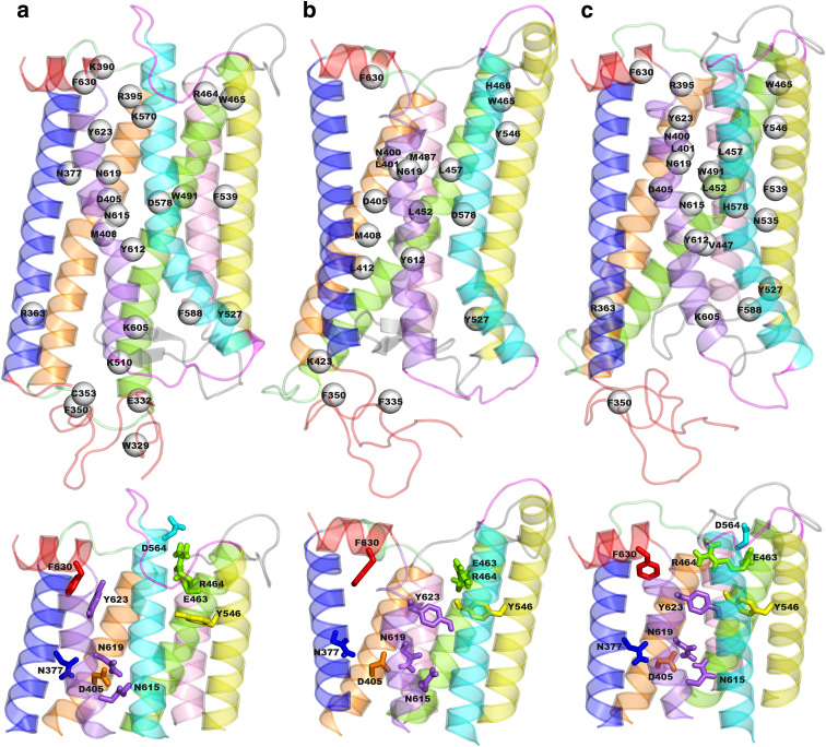
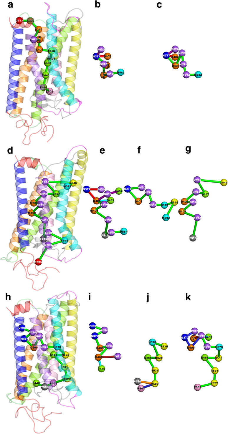
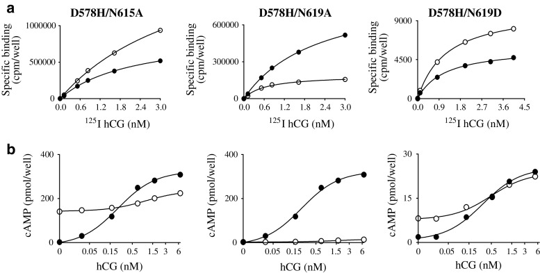
The luteinizing hormone receptor (LHR) is a G protein-coupled receptor (GPCR) particularly susceptible to spontaneous pathogenic gain-of-function mutations. Protein structure network (PSN) analysis on wild-type LHR and two constitutively active mutants, combined with in vitro mutational analysis, served to identify key amino acids that are part of the regulatory network responsible for propagating communication between the extracellular and intracellular poles of the receptor. Highly conserved amino acids in the rhodopsin family GPCRs participate in the protein structural stability as network hubs in both the inactive and active states. Moreover, they behave as the most recurrent nodes in the communication paths between the extracellular and intracellular sides in both functional states with emphasis on the active one. In this respect, non-conservative loss-of-function mutations of these amino acids is expected to impair the most relevant way of communication between activating mutation sites or hormone-binding domain and G protein recognition regions.


Figures

Fig. 1
Fig. 1
Hub distribution and details of interactions involving highly conserved amino acids. Top The hub residues in the wild-type (a), in D564(6.30)G (b), and D578(6.44)H (c) are shown as white spheres centered on the Cα-atoms on the average minimized structures. Only hubs characterized by frequencies higher than 30% are shown. The receptor structures are seen in a direction parallel to the putative membrane surface, the cytosolic side being on top. The different receptor portions are colored as follows: helices 1, 2, 3, 4, 5, 6, 7 and 8 are blue, orange, green, pink, yellow, cyan, violet; and red; the N-ter is red, IL1 and EL1 are lime, IL2 and EL2 are grey, and IL3 and EL3 are magenta. Bottom The cytosolic halves of the wild-type (a), D564(6.30)G (b) and D578(6.44)H (c) are shown according to the same view as on top. Details of the interactions made by the highly conserved amino acids are shown as sticks
Fig. 2
Fig. 2
Representative paths in the wild-type (top), D564(6.30)G (middle) and D578(6.44)H (bottom). The receptor structures are seen in a direction parallel to the putative membrane surface, the cytosolic side being on top. The longest paths with the highest relative frequency amongst the total number of paths (i.e., paths with frequency ≥ 10% and length ≥ 3) are shown on the bundle. In contrast, paths selected among those characterized by frequency ≥ 30% and length ≥ 5, i.e., most frequent paths, are shown outside the bundle. The amino acid composition of the longest path is reported below, whereas those of the most frequent paths are listed in Tables S1–S3. In general, nodes are represented by spheres centered on the Cα-atoms and connections between nodes are represented by green sticks. In those cases in which multiple paths are shown, green sticks indicate the shared portion of the paths. Here, the identification number of the selected paths together with their respective frequency are reported. In this respect, those paths graphically shown for the wild-type are: a L502F444F448L452W491T456L401M398Y623R395K390D633 the longest path with the relative highest frequency (13.79%); b paths 1, (44.55%; red stick) and 5 (33.88%; purple stick); and c paths 2 (41.37%; red stick) and 3 (39.05%; purple stick). Those paths graphically shown for D564(6.30)G are: d F335K605F588L608F515Y612M408S616D405N619L457Y623Y546M571 the longest path with the relative highest frequency (11.07%); e paths 8 (47.2%; red sticks), 9 (43.72%; purple sticks) and 10 (43.28%; cyan stick); f path 23 (35.60%); and g path 46 (30.33%). Those paths graphically shown for D578(6.44)H are: h S440Y508P501Y527I531E451N535H578N615N619D405N377P620V380 the longest path with the relative highest frequency (11.58%); i paths 1 (56.46%; red sticks) and 4 (46.85%; cyan stick); j paths 6 (46.09%; blue stick) and 9 (43.16%; red stick); and k paths 24 (35.77%; purple stick), 29 (33.78%; cyan sticks), and 38 (31.26%; blue sticks)
Fig. 3
Fig. 3
Representative saturation binding curves a and hCG-mediated cAMP responses b for wild-type LHR (closed circles) and three double mutants (open circles). D578(6.44)H paired with N615(7.45)A, N619(7.49)A and N619(7.49)D (comparable figures are given in the Supplement)

References

    1. Ascoli M, Puett D. The gonadotropin hormones and their receptors. In: Strauss JF III, Barbieri RR, editors. Yen and Jaffee’s reproductive endocrinology. 6. Philadelphia: Elsevier; 2009. pp. 33–55.
    1. Li J, Edwards PC, Burghammer M, Villa C, Schertler GF. Structure of bovine rhodopsin in a trigonal crystal form. J Mol Biol. 2004;343:1409–1438. doi: 10.1016/j.jmb.2004.08.090. - DOI - PubMed
    1. Okada T, Sugihara M, Bondar AN, Elstner M, Entel P, Buss V. The retinal conformation and its environment in rhodopsin in light of a new 2.2 A crystal structure. J Mol Biol. 2004;342:571–583. doi: 10.1016/j.jmb.2004.07.044. - DOI - PubMed
    1. Park JH, Scheerer P, Hofmann KP, Choe HW, Ernst OP. Crystal structure of the ligand-free G-protein-coupled receptor opsin. Nature. 2008;454:183–187. doi: 10.1038/nature07063. - DOI - PubMed
    1. Warne T, Serrano-Vega MJ, Baker JG, Moukhametzianov R, Edwards PC, Henderson R, Leslie AG, Tate CG, Schertler GF. Structure of a beta1-adrenergic G-protein-coupled receptor. Nature. 2008;454:486–491. doi: 10.1038/nature07101. - DOI - PMC - PubMed

Publication types

LinkOut - more resources