Review
. 2010 May-Jun;2(3):305-323.
doi: 10.1002/wsbm.59.

Building the developmental oculome: systems biology in vertebrate eye development and disease

Affiliations
Review

Building the developmental oculome: systems biology in vertebrate eye development and disease

Salil A Lachke et al. Wiley Interdiscip Rev Syst Biol Med. 2010 May-Jun.

Abstract

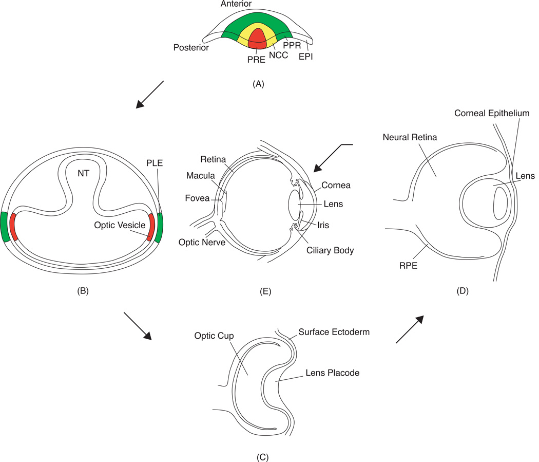
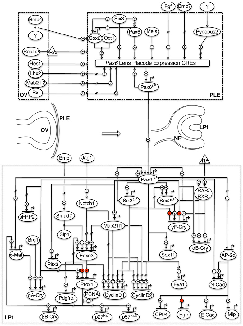
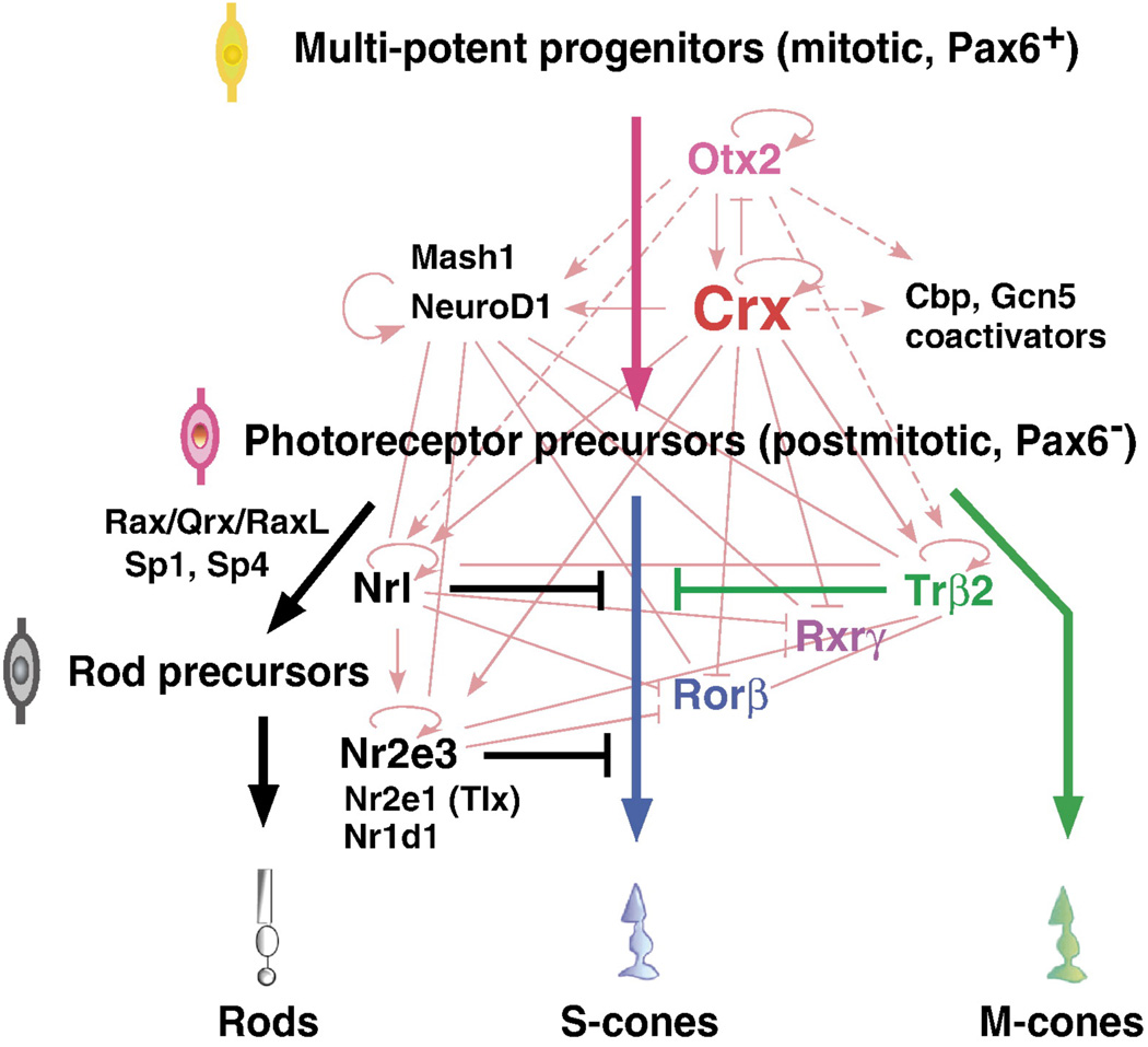
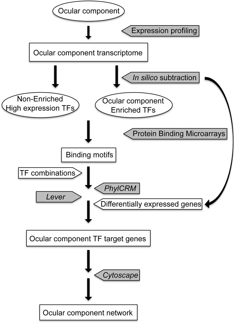
The vertebrate eye is a sophisticated multicomponent organ that has been actively studied for over a century, resulting in the identification of the major embryonic and molecular events involved in its complex developmental program. Data gathered so far provides sufficient information to construct a rudimentary network of the various signaling molecules, transcription factors, and their targets for several key stages of this process. With the advent of genomic technologies, there has been a rapid expansion in our ability to collect and process biological information, and the use of systems-level approaches to study specific aspects of vertebrate eye development has already commenced. This is beginning to result in the definition of the dynamic developmental networks that operate in ocular tissues, and the interactions of such networks between coordinately developing ocular tissues. Such an integrative understanding of the eye by a comprehensive systems-level analysis can be termed the 'oculome', and that of serial developmental stages of the eye as it transits from its initiation to a fully formed functional organ represents the 'developmental oculome'. Construction of the developmental oculome will allow novel mechanistic insights that are essential for organ regeneration-based therapeutic applications, and the generation of computational models for eye disease states to predict the effects of drugs. This review discusses our present understanding of two of the individual components of the developing vertebrate eye--the lens and retina--at both the molecular and systems levels, and outlines the directions and tools required for construction of the developmental oculome.


Figures

Fig. 1
Fig. 1
Vertebrate ocular development. (A) In late gastrulation, the anterior neural plate contains the presumptive retinal ectoderm (PRE) surrounded by neural crest cells (NCC), pre-placodal region (PRR), and the epidermis (EPI). (B) As the neural tube (NT) closes, the diencephalon bilaterally develops into the optic vesicles, which contact the presumptive lens ectoderm (PLE) on either side. (C) Co-ordinate signaling leads to induction of the PLE to form the lens placode, while the optic vesicle invaginates to form the optic cup. (D) The lens placode invaginates and forms the lens pit that detaches from the surface ectoderm to form the lens vesicle. At this stage the optic cup starts to differentiate into the neural retina and the retinal pigment epithelium (RPE). (E) The adult vertebrate eye thus contains multiple tissue compartments. It is important to note that in fish and frogs, the lens placode does not form a hollow lens vesicle, but instead develops into a solid aggregate of cells.
Fig. 2
Fig. 2
The molecular circuitry of early mammalian lens development. Numerous signals from the optic vesicle (OV) along with other molecules converge on multiple cis-regulatory elements (CREs) to induce the placodal expression of Pax6 (Pax6LP) in the presumptive lens ectoderm (PLE). As the PLE develops into the lens pit (LPt), Pax6LP turns on the circuitry necessary for lens formation. Pax6LP, Six3LP and Sox2LP indicate expression of these genes in stages following lens placode induction. Hatches in lines indicate that the interaction may involve intermediate steps.
Fig. 3
Fig. 3
Transcription regulatory network (TRN) for retinal photoreceptor cell development. Multipotent, photoreceptor precursor cells differentiate into individual photoreceptor cell types via three separate pathways. Thin lines indicate protein-promoter interactions; solid lines indicate published data and dotted lines are from unpublished data of Hennig et al 2008 [145]. Reprinted from Brain Research 1192, Hennig, A.K., Peng, G-H., Chen, S., Regulation of photoreceptor gene expression by Crx-associated transcription factor network, 114–133, (2008) with permission from Elsevier.
Fig. 4
Fig. 4
Strategy outlining the construction of networks for specific ocular compartments using gene expression profiling and computational tools. Experimental steps are indicated in gray boxes and names of computational software are italicized.

Similar articles

Cited by

References

    1. McCulloch AD. Modeling the human cardiome in silico. J Nucl Cardiol. 2000;7:496–499. - PubMed
    1. McCulloch AD, Paternostro G. Cardiac systems biology. Ann N Y Acad Sci. 2005;1047:283–295. - PubMed
    1. Srivastava R, Varner J. Emerging technologies: systems biology. Biotechnol Prog. 2007;23:24–27. - PubMed
    1. Monte J, Sakurai H, Bush K, Nigam S. The developmental nephrome: systems biology in the developing kidney. Current Opinion in Nephrology and Hypertension. 2007;16:3–9. - PubMed
    1. Busser BW, Bulyk ML, Michelson AM. Toward a systems-level understanding of developmental regulatory networks. Curr Opin Genet Dev. 2008;18:521–529. - PMC - PubMed

Publication types